Review
. 2005 Nov;207(5):637-53.
doi: 10.1111/j.1469-7580.2005.00475.x.

Growth of the normal skull vault and its alteration in craniosynostosis: insights from human genetics and experimental studies

Affiliations
Review

Growth of the normal skull vault and its alteration in craniosynostosis: insights from human genetics and experimental studies

Gillian M Morriss-Kay et al. J Anat. 2005 Nov.

Abstract

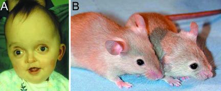
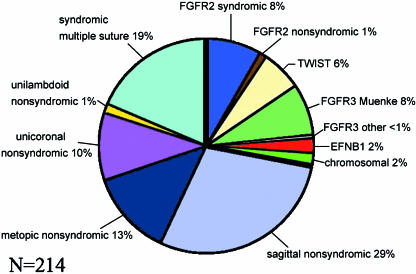
The mammalian skull vault is constructed principally from five bones: the paired frontals and parietals, and the unpaired interparietal. These bones abut at sutures, where most growth of the skull vault takes place. Sutural growth involves maintenance of a population of proliferating osteoprogenitor cells which differentiate into bone matrix-secreting osteoblasts. Sustained function of the sutures as growth centres is essential for continuous expansion of the skull vault to accommodate the growing brain. Craniosynostosis, the premature fusion of the cranial sutures, occurs in 1 in 2500 children and often presents challenging clinical problems. Until a dozen years ago, little was known about the causes of craniosynostosis but the discovery of mutations in the MSX2, FGFR1, FGFR2, FGFR3, TWIST1 and EFNB1 genes in both syndromic and non-syndromic cases has led to considerable insights into the aetiology, classification and developmental pathology of these disorders. Investigations of the biological roles of these genes in cranial development and growth have been carried out in normal and mutant mice, elucidating their individual and interdependent roles in normal sutures and in sutures undergoing synostosis. Mouse studies have also revealed a significant correspondence between the neural crest-mesoderm boundary in the early embryonic head and the position of cranial sutures, suggesting roles for tissue interaction in suture formation, including initiation of the signalling system that characterizes the functionally active suture.


Figures

Fig. 1
Fig. 1
Bones and sutures of the newborn human skull (A,B) and late fetal mouse skulls (stained with Alizarin red for bone and Alcian blue for cartilage) (C,D). (A,B) Lateral and vertex views; some bone has been removed to show the teeth. (C) E17.5 skull, lateral view; (D) E18.5 skull, vertex view (skull base removed for clarity). Membrane bones: al, alisphenoid; f, frontal; ip, interparietal; n, nasal; p, parietal; sq, squamosal. The membrane bones of the upper and lower jaws (maxilla and mandible) are unlabelled. Endochondral bones: eo, exoccipital; pt, petrous temporal; so, supraoccipital. Sutures: cs, coronal; ls, lambdoid; ms, metopic (interfrontal); fm, foramen magnum; ss, sagittal; af, anterior fontanelle.
Fig. 10
Fig. 10
Crouzon syndrome. (A) Characteristic flattened midface and proptosis due to shallow orbits; the domed skull shape compensates for premature loss of the coronal suture (see Fig. 2). (B) Fgfr2C342Y mouse (right) showing shortened face, proptosis and domed skull vault compared with the wild-type head.
Fig. 2
Fig. 2
Three-dimensional computer tomography scans showing sagittal synostosis in a child of approximately 4 years of age (A,B) and coronal synostosis in a young baby (C,D). Decreased growth in the plane of growth of the sagittal suture is compensated by increased growth in the fronto-occipital plane (A,B); decreased growth in the plane of growth of the coronal suture (C) is compensated for by increased growth in breadth, and the metopic suture is widely open (D). Arrows: open (functional) sutures; asterisk: position of fused sagittal suture (A) and coronal suture (C).
Fig. 3
Fig. 3
Organization of the neural folds and migration of the neural crest cells in mouse embryos. (A,H) Scanning electron micrographs; (B–G) X-gal-stained Wnt1-Cre/R26R embryos. A, four-somite (s)-stage scanning electron micrograph, showing preotic (arrow) and otic (arrowhead) sulci, prorhombomeres (A–C) (lettered) the occipital region (oc) and the primitive streak region with Henson's node (hn); B, 5s embryo, frontal view, showing emigrating neural crest cells (nc); (C–G) embryos of somite stages as indicated, showing migration of neural crest cells from the neural folds anterior to the preotic sulcus (arrowed) into the frontonasal and first arch regions; in the 23s embryo, the neural crest–mesoderm boundary is clearly defined (arrowheads). (H) 18s-stage embryo, bisected to show rhombomeres (numbered) and position of former preotic and otic sulci (arrow and arrowhead). A–C, Prorhombomeres; d, diencephalon; e, eye; fb, forebrain; fn, frontonasal mesenchyme; ht, heart; h, hyoid neural crest cell population; m, mandibular part of first branchial arch; mb, midbrain; n, notochord; p, pharynx; t, telencephalon; V, trigeminal ganglion crest cells; va, vagal crest cells. Images B, C, E and F were previously published in Jiang et al. (2002). Scale bars: 200 µm.
Fig. 4
Fig. 4
Neural crest and mesodermal contributions to the mouse head at E17.5. (A)Lateral view of a whole Wnt1-Cre/R26R head stained with Alizarin red to show mineralized bone and X-gal (blue–green) to reveal neural crest-derived tissues, including the meningeal covering of the cerebral hemispheres (arrowheads); neural crest-derived tissue extends into the sagittal suture (black arrow) and there is a separate hindbrain-derived patch in the interparietal region (white arrows). (B) Section of the coronal suture stained with X-gal and fast red, showing the neural crest-derived frontal bone (f) and the mesoderm-derived parietal bone (dashed outline) overlying the meninges (m) of the cerebral hemisphere (ch). (C) Diagram showing the neural crest-derived (blue) and mesodermal (red) contributions to the skull vault at E17.5. Modified from images in Jiang et al. (2002). bo, basioccipital; e, eye; m, meninges; pn, pinna of ear; s, skin. Other labels as Fig. 1. Scale bars: A, 1 mm; B, 100 µm.
Fig. 5
Fig. 5
Structure of fibroblast growth factor receptor proteins (types 1–3), showing the position of some of the common mutations causing craniosynostosis, and (FGFR3 only) some of the mutations affecting long-bone growth. Each receptor has three immunoglobulin-like domains (Ig) whose structure is maintained by disulphide bonds (s–s); TM, transmembrane domain; TK1,2, tyrosine kinase domain.
Fig. 6
Fig. 6
Craniofrontonasal dysplasia due to EFNB1 mutation (A,B) and wild-type mouse embryos (C–F) showing (arrowed) the correspondence between the frontonasal neural crest–mesoderm boundary in Wnt1-Cre/R26R embryos (C,D) and Efnb1 expression (E,F) in whole heads (C,E) and sections (D,F). (C,E,F) E10.5; (D) E12. ch, cerebral hemisphere; other labels as Fig. 1. C, modified from Jiang et al. (2002); A, E and F were previously published in Twigg et al. (2004). Scale bars: C, E: 1 mm; D, F: 200 µm.
Fig. 7
Fig. 7
Genetic epidemiology of craniosynostosis, based on a prospective series of 214 patients born between 1993 and 2005 inclusive, and screened for mutations in the entire coding region of FGFR1, FGFR2, FGFR3, TWIST1 and EFNB1. A specific molecular diagnosis was made in 60 (28%) of cases: this is represented as the segment between the thick black lines.
Fig. 8
Fig. 8
Diagram illustrating the alternative use of either exon IIIb or IIIc to form alternative splice forms of FGFR receptors, in this case FGFR2b and FGFR2c, respectively.
Fig. 9
Fig. 9
The relationship between Fgfr expression and the progression from proliferating osteoprogenitor cells to differentiating osteoblasts at the edge of a bone in the mouse coronal suture. (A) Osteoblasts (blue) express Fgfr1 and secrete bone matrix proteins (light blue) and FGF (pink); FGF diffuses into the region of proliferating osteoprogenitor cells (green). (B) Hypothetical scheme suggesting that a threshold of FGF concentration effects the change in gene expression from Fgfr2 to Fgfr1 and the change in cell behaviour from proliferation to differentiation.

References

    1. Anderson J, Burns HD, Enriquez-Harris P, Wilkie AOM, Heath JK. Apert syndrome mutations in fibroblast growth factor receptor 2 exhibit increased affinity for FGF ligand. Hum Mol Genet. 1998;7:1475–1483. - PubMed
    1. Arman E, Haffner-Krausz R, Chen Y, Heath JK, Lonai P. Targeted disruption of FGFR2 suggests a role for FGF signalling in pre-gastrulation mammalian development. Proc Natl Acad Sci USA. 1998;95:5082–5087. - PMC - PubMed
    1. Beer HD, Vindevoghel L, Gait MJ, et al. Fibroblast growth factor (FGF) receptor 1-IIIb is a naturally occurring functional receptor for FGfs that is a naturally occurring functional receptor for FGfs that is preferentially expressed in the skin and brain. J Biol Chem. 2000;275:16091–16097. - PubMed
    1. Beiman M, Shilo BZ, Volk T. Heartless, a Drosophila FGF receptor homolog, is essential for cell migration and establishment of several mesodermal lineages. Genes Dev. 1996;10:2993–3002. - PubMed
    1. Bellus GA, Gaudenz K, Zackai EH, et al. Identical mutations in three different fibroblast growth factor receptor genes in autosomal dominant craniosynostosis syndromes. Nature Genet. 1996;14:174–176. - PubMed

Publication types

LinkOut - more resources