

Three-dimensional analysis of mandibular growth and tooth eruption
- PMID: 16313399
- PMCID: PMC1571566
- DOI: 10.1111/j.1469-7580.2005.00479.x
Three-dimensional analysis of mandibular growth and tooth eruption
Abstract
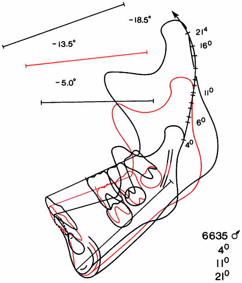
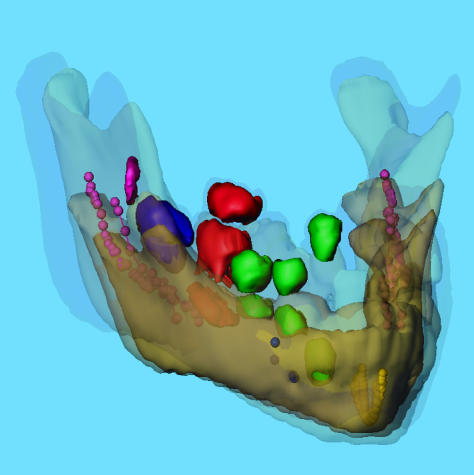
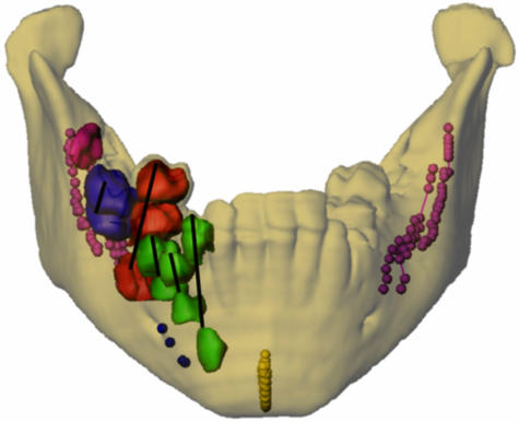
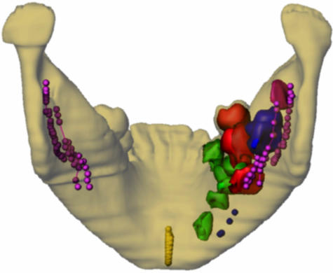
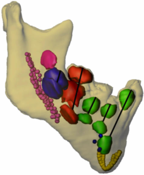
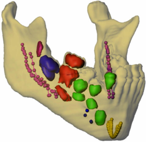
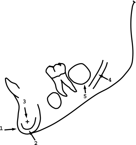
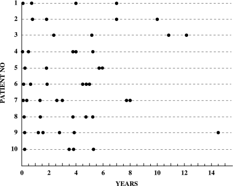
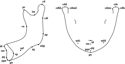
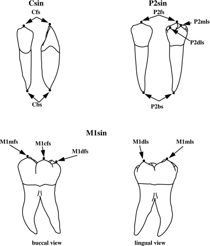
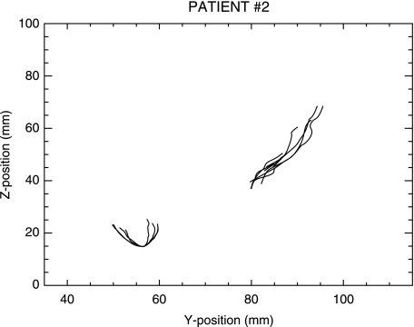
Normal and abnormal jaw growth and tooth eruption are topics of great importance for several dental and medical disciplines. Thus far, clinical studies on these topics have used two-dimensional (2D) radiographic techniques. The purpose of the present study was to analyse normal mandibular growth and tooth eruption in three dimensions based on computer tomography (CT) scans, extending the principles of mandibular growth analysis proposed by Björk in 1969 from two to three dimensions. As longitudinal CT data from normal children are not available (for ethical reasons), CT data from children with Apert syndrome were employed, because it has been shown that the mandible in Apert syndrome is unaffected by the malformation, and these children often have several craniofacial CT scans performed during childhood for planning of cranial and midface surgery and for follow-up after surgery. A total of 49 datasets from ten children with Apert syndrome were available for study. The number of datasets from each individual ranged from three to seven. The first CT scan in each of the ten series was carried out before 1 year of age, and the ages for the 49 scans ranged from 1 week to 14.5 years. The mandible and the teeth were segmented and iso-surfaces generated. Landmarks were placed on the surface of the mandible, along the mandibular canals, the inner contour of the cortical plate at the lower border of the symphysis menti, and on the teeth. Superimposition of the mandibles in the longitudinal series was performed using the symphysis menti and the mandibular canals as suggested by Björk. The study supported the findings of stability of the symphysis menti and the mandibular canals as seen in profile view previously reported by Björk & Skieller in 1983. However, the mandibular canals were, actually, relocated laterally during growth. Furthermore, the position of tooth buds remained relatively stable inside the jaw until root formation started. Eruption paths of canines and premolars were vertical, whereas molars erupted in a lingual direction. The 3D method would seem to offer new insight into jaw growth and tooth eruption, but further studies are needed.
Figures
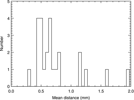
 , between the first and the second digitization (N = 25).
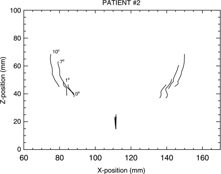
References
-
- Abbott AH, Netherway DJ, David DJ, Brown T. Application and comparison of techniques for three dimensional analysis of craniofacial anomalies. J Craniofac Surg. 1990;1:119–134. - PubMed
-
- Abbott AH, Netherway DJ, Niemann DB, et al. CT-determined intracranial volume for a normal population. J Craniofac Surg. 2000;11:211–223. - PubMed
-
- AnalyzeDirect. Analyze. 1999. Version 3.1. ©1999-2001 AnalyzeDirect and ©1986-2001 Mayo Foundation for Medical Education and Research.
-
- Anamedic. Mvox. 1999. version 1.41. ©1999 Anamedic & Morten Bro-Nielsen.
-
- Andresen PR, Bookstein FL, Conradsen K, Ersbøll BK, Marsh JL, Kreiborg S. Surface-bounded growth modelling applied to human mandibles. IEEE Trans Med Imaging. 2000;19:1053–1063. - PubMed
Publication types
MeSH terms
LinkOut - more resources
Full Text Sources
Medical
Research Materials
