. 2005 Dec;58(5):1303-21.
doi: 10.1111/j.1365-2958.2005.04905.x.

Characterization of point mutations in the cdtA gene of the cytolethal distending toxin of Actinobacillus actinomycetemcomitans

Affiliations

Characterization of point mutations in the cdtA gene of the cytolethal distending toxin of Actinobacillus actinomycetemcomitans

Linsen Cao et al. Mol Microbiol. 2005 Dec.

Abstract

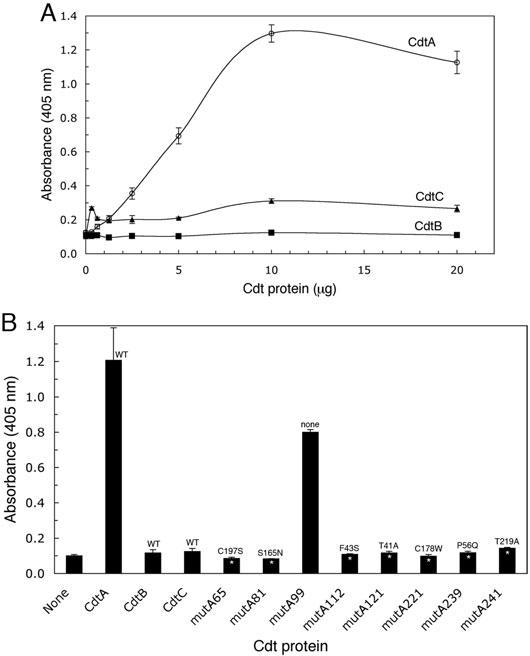
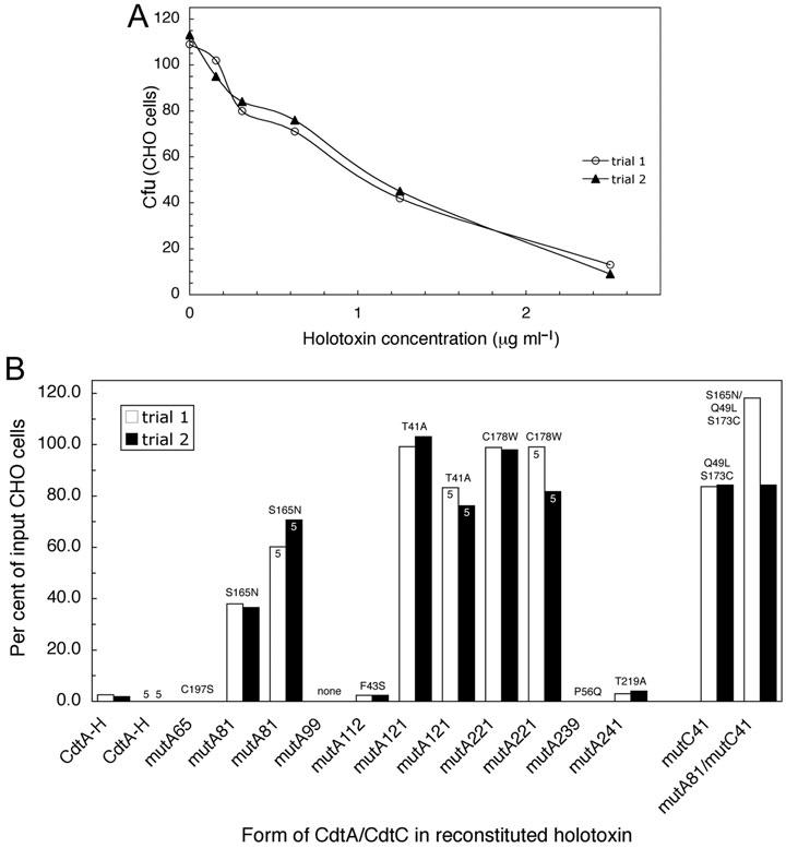
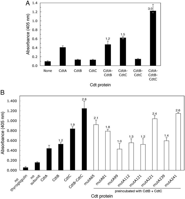
The Cdt is a family of gram-negative bacterial toxins that typically arrest eukaryotic cells in the G0/G1 or G2/M phase of the cell cycle. The toxin is a heterotrimer composed of the cdtA, cdtB and cdtC gene products. Although it has been shown that the CdtA protein subunit binds to cells in culture and in an enzyme-linked immunosorbent assay (CELISA) the precise mechanisms by which CdtA interacts with CdtB and CdtC has not yet been clarified. In this study we employed a random mutagenesis strategy to construct a library of point mutations in cdtA to assess the contribution of individual amino acids to binding activity and to the ability of the subunit to form biologically active holotoxin. Single unique amino acid substitutions in seven CdtA mutants resulted in reduced binding of the purified recombinant protein to Chinese hamster ovary cells and loss of binding to the fucose-containing glycoprotein, thyroglobulin. These mutations clustered at the 5'- and 3'-ends of the cdtA gene resulting in amino acid substitutions that resided outside of the aromatic patch region and a conserved region in CdtA homologues. Three of the amino acid substitutions, at positions S165N (mutA81), T41A (mutA121) and C178W (mutA221) resulted in gene products that formed holotoxin complexes that exhibited a 60% reduction (mutA81) or loss (mutA121, mutA221) of proliferation inhibition. A similar pattern was observed when these mutant holotoxins were tested for their ability to induce cell cycle arrest and to convert supercoiled DNA to relaxed and linear forms in vitro. The mutations in mutA81 and mutA221 disrupted holotoxin formation. The positions of the amino acid substitutions were mapped in the Haemophilus ducreyi Cdt crystal structure providing some insight into structure and function.


Figures

Fig. 1
Fig. 1
Effect of point mutations on CdtA binding to CHO cells. Affinity-purified CdtA mutant proteins were incubated with CHO cells in a CELISA. A. Saturation curves of wild-type recombinant CdtA-His6, CdtB-His6, CdtC-His6 and total soluble protein from E. coli BL-21(DE3) (pET15b) in a CELISA. Bound Cdt protein subunit was detected with anti-HisTag monoclonal antibody (1:3000 dilution) and anti-mouse IgG horseradish peroxidase conjugate (1:3000 dilution). B. Recombinant CdtA-His6 mutant proteins (1 μg well−1) were incubated with CHO cells in a CELISA. Bound protein was detected as in the experiment in A and compared with wells containing wild-type CdtA-His6, CdtB-His6 and CdtC-His6. Amino acid substitutions are shown for each mutant. All samples were run in triplicate. Statistically significant differences between the mutants and wild-type CdtA-His6 are marked with asterisks (P < 0.005*, P < 0.01**). Results are representative of three trials.
Fig. 2
Fig. 2
Effect of point mutations on CdtA binding to thyroglobulin. A. Saturation curves of the binding of wild-type recombinant CdtA-His6, CdtB-His6, CdtC-His6 to thyroglobulin-coated wells in ELISA. Bound protein was detected as described in the legend to Fig. 1. B. Affinity-purified recombinant CdtA-His6 mutant proteins (10 μg well−1) were added to wells coated with thyroglobulin. Bound protein was detected as in the experiment in A and compared with wells containing 10 μg well−1 of wild-type CdtA-His6, CdtB-His6 and CdtC-His6. Amino acid substitutions are designated for each mutant. All samples were run in triplicate. Statistically significant differences between the absorbance values for mutant and wild-type CdtA-His6 are marked with an asterisk (P < 0.005). Results are typical of three trials.
Fig. 3
Fig. 3
Effect of point mutations in CdtA on the proliferation of CHO cells treated with reconstituted holotoxin. A. Wild-type recombinant CdtA-His6, CdtB-His6 and CdtC-His6 (1:1:1 by weight) were preincubated in folding buffer and increasing concentrations of the mixture was added to CHO cell cultures. Cell colonies were stained and counted after 6 days and the data expressed as cfu. B. Holotoxin was reconstituted as in the experiment in A except that recombinant CdtA-His6 mutant proteins were substituted for wild-type CdtA-His6. In some samples a CdtC-His6 mutant (mutC41) was substituted for wild-type CdtC-His6. Amino acid substitutions are shown for each mutant. MutC41 contained two amino acid substitutions. CdtA-H designates holotoxin reconstituted with wild-type CdtA-His6. All samples were run twice in single wells. Twice the amount of CdtA-His6 protein (5 μg ml−1) was used to treat some cultures. Cfu were obtained as in A and the data is expressed as the percent of input CHO cells.
Fig. 4
Fig. 4
Effect of point mutations in the cdtA gene on the cell cycle of CHO cells treated with reconstituted holotoxin. A. CHO cell cultures (1 × 10⁶) were treated with 0, 2.5, 5.0, 10.0 and 25.0 μg of total holotoxin protein ml−1. Holotoxin was reconstituted from wild-type CdtA-His6, CdtB-His6 and CdtC-His6 (1:1:1) for 36 h post intoxication. DNA profiles obtained by FACS analysis of propidium iodide stained nuclei are shown (solid line). G0/G1 (2n) and G2/M (4n) (solid filled) and S (diagonal line filled) peaks where determined by computer analysis. B. Dose–response curve of the percentage of cells having a 4n DNA content after treatment with each holotoxin concentration. C. CHO cell cultures were treated as in A except that 10 μg of total mutant CdtA or CdtC holotoxin protein ml−1 was used. Panels a, mutA65; b, mutA81; c, mutA99; d, mutA112; e, mutA121; f, mutA221; g, mutA239; h, mutA241; i, mutC41. Results are representative of two experiments.
Fig. 5
Fig. 5
Effect of point mutations in the cdtA gene on the in vitro DNA nicking activity of reconstituted holotoxin. A. Wild-type Cdt subunit proteins alone and in various combinations were incubated with pBluescript II SK(+) DNA under conditions optimized for CdtB-His6 nuclease activity (Experimental procedures). B. Holotoxin prepared with protein from each of the CdtA-His6 mutants and from mutC41 was incubated with plasmid DNA as in A. Samples were examined on 0.8% agarose gels stained with ethidium bromide. Supercoiled (S), linear (L) and relaxed (R) forms of DNA are labelled. Results are representative of three experiments.
Fig. 6
Fig. 6
Competitive binding of mutant CdtA-His6 proteins to CdtB-His6 and CdtC-His6. A. Subunit, heterodimer and heterotrimer binding to thyroglobulin. ELISA plates were coated with thyroglobulin followed by the addition of individual or a combination of wild-type subunit proteins. B. Competition assay. ELISA plates were coated with thyroglobulin followed by 4 μg well−1 of wild-type CdtA-His6. Individual wild-type CdtB-His6 and CdtC-His6 subunit proteins, a preincubated mixture of CdtB-His6 with CdtC-His6 and preincubated mixtures of the mutant CdtA-His6 proteins with wild-type CdtB-His6 and CdtC-His6 were added to the plates. The plates were developed and the absorbance determined as described in the legend to Fig. 1. All samples were run in triplicate. Absorbance ratios based on the binding of wild-type CdtA-His6 to thyroglobulin were calculated as described in Experimental procedures. A statistically significant increase in the absorbance values for the homodimers and heterotrimer relative to that of wild-type CdtA-His6 are marked with an asterisks (P < 0.005*, P < 0.01**). Results are representative of two experiments.
Fig. 7
Fig. 7
Effect of point mutations in the cdtA gene on the reconstitution of holotoxin. Individual wild-type subunit proteins and heterotrimers composed of preincubated mixtures of either wild-type or mutant CdtA-His6 proteins were dialysed for 48 h. The protein composition of the material in the dialysis bag, before and after dialysis, was then examined by Western blotting as described in the Experimental procedures. BD, before dialysis; AD, after dialysis.
Fig. 8
Fig. 8
Alignment of A. actinomycetemcomitans recombinant CdtA-His6 (pJDA9), H. ducreyi CdtA (GenBank Accession number U53215) and CdtA-His6 mutant deduced amino acid sequences (Table 3). Identical residues are boxed. The positions of the four amino acid substitutions in the aromatic patch (ap) mutant described in reference 24 are marked. Amino acid resides 18–56 were disordered in the crystal structure (NeŠić et al., 2004). The arrow marks the amino-terminal end of the 17–18 kDa form of CdtA identified in several studies (Frisk et al., 2001; Saiki et al., 2004; Shenker et al., 2004). The solid line between the two sequences marks the region conserved among the various CdtA proteins (Lee et al., 2003).
Fig. 9
Fig. 9
Location of the amino acid substitutions of the CdtA-His6 mutant proteins in the crystal structure of the H. ducreyi Cdt. The coordinates (Protein Data Bank Accession number 1SR4) for the crystal structure (NeŠić et al., 2004) were computer modelled. A–C. Orientation of the CdtA, CdtC and CdtB subunits respectively. The amino-(N) and carboxy-(C) terminal residues in CdtA and CdtC and the theoretical active site in CdtB are labelled. D. Locations of the seven unique amino acid substitutions (black text) in the A. actinomycetemcomitans recombinant CdtA-His6 mutants that affected binding are shown at the positions of the corresponding residues in the H. ducreyi sequence. The positions of the three substitutions that resulted in a non-cytotoxic phenotype are underlined. The CdtA used to crystalize the H. ducreyi Cdt contained residues 18–223 to eliminate a putative secretory signal sequence (NeŠić et al., 2004). Residues 18–56 were disordered in the crystal structure. The two amino acid substitutions in the CdtC-His6 mutant (mutC41) are shown in red. E. Location of the four amino acid substitutions in the aromatic patch (AP) region mutant of NeŠić et al. (2004). F. Locations of the four cysteine residues in CdtA. These cysteines are conserved in the A. actinomycetemcomitans and H. ducreyi CdtA.

References

    1. Akifusa S, Poole S, Lewthwaite J, Henderson B, Nair SP. Recombinant Actinobacillus actinomycetemcomitans cytolethal distending toxin proteins are required to interact to inhibit human cell cycle progression and to stimulate human leukocyte cytokine synthesis. Infect Immun. 2001;69:5925–5930. - PMC - PubMed
    1. Akifusa S, Heywood W, Nair SP, Stenbeck G, Henderson B. Mechanism of internalization of the cytolethal distending toxin of Actinobacillus actinomycetemcomitans. Microbiology. 2005;151:1395–1402. - PubMed
    1. Altschul SF, Madden TL, Schäffer AA, Zhang J, Zhang Z, Miller W, Lipman DJ. Gapped blast and psi-blast: a new generation of protein database search programs. Nucleic Acids Res. 1997;25:3389–3402. - PMC - PubMed
    1. Cope LD, Lumbley S, Latimer JL, Klesney-Tait J, Stevens MK, Johnson LS, et al. A diffusible cytotoxin of Haemophilus ducreyi. Proc Natl Acad Sci USA. 1997;94:4056–4061. - PMC - PubMed
    1. Cortes-Bratti X, Frisan T, Thelestam M. The cytolethal distending toxins induce DNA damage and cell cycle arrest. Toxicon. 2001a;39:1729–1736. - PubMed

Publication types

MeSH terms