. 2005 Nov 24;33(20):6555-65.
doi: 10.1093/nar/gki964. Print 2005.

Human Bex2 interacts with LMO2 and regulates the transcriptional activity of a novel DNA-binding complex

Affiliations

Human Bex2 interacts with LMO2 and regulates the transcriptional activity of a novel DNA-binding complex

Chunyu Han et al. Nucleic Acids Res. .

Abstract

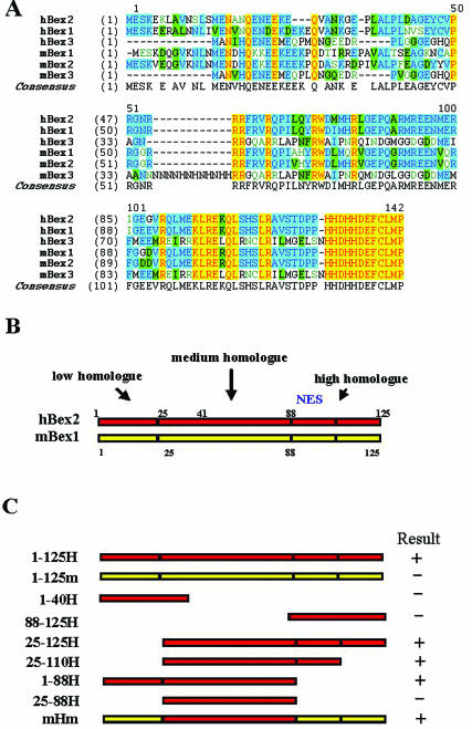
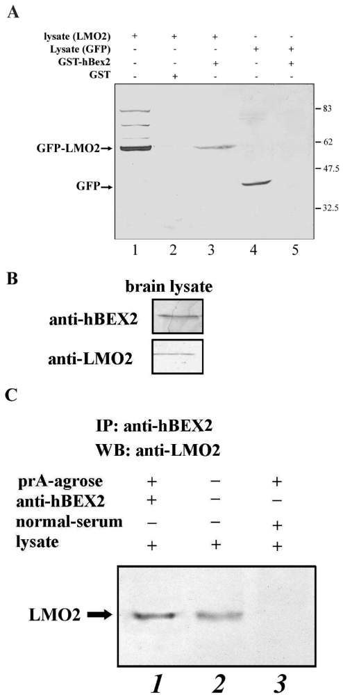
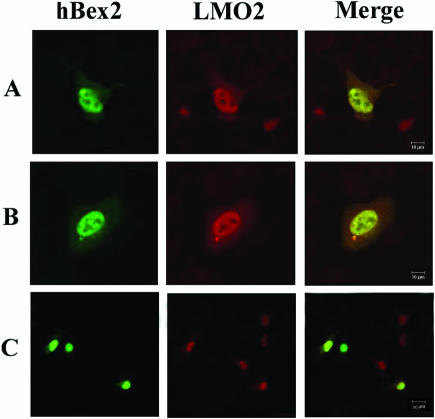
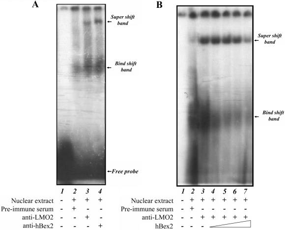
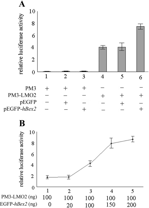
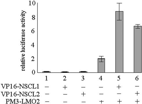
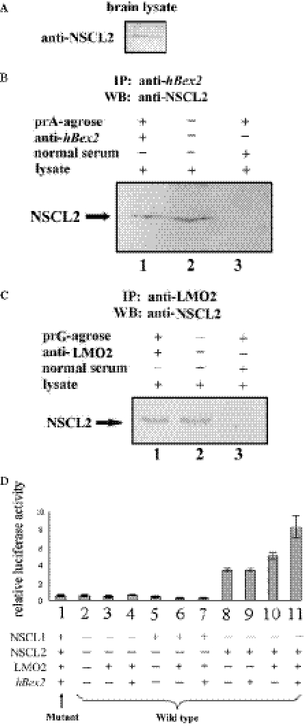
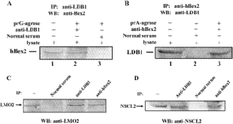
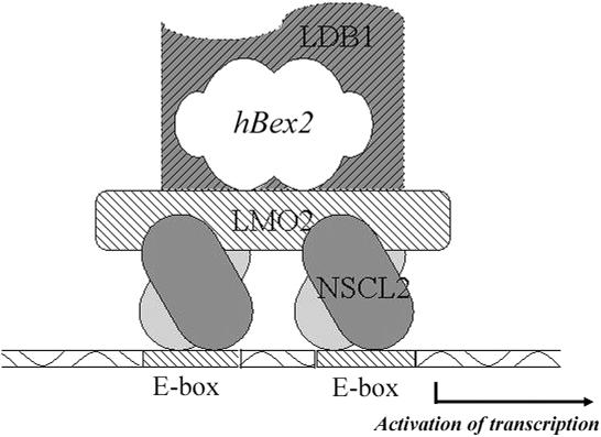
Human Bex2 (brain expressed X-linked, hBex2) is highly expressed in the embryonic brain, but its function remains unknown. We have identified that LMO2, a LIM-domain containing transcriptional factor, specifically interacts with hBex2 but not with mouse Bex1 and Bex2. The interaction was confirmed both by pull-down with GST-hBex2 and by coimmunoprecipitation assays in vivo. Using electrophoretic mobility shift assay, we have demonstrated the physical interaction of hBex2 and LMO2 as part of a DNA-binding protein complex. We have also shown that hBex2 can enhance the transcriptional activity of LMO2 in vivo. Furthermore, using mammalian two-hybrid analysis, we have identified a neuronal bHLH protein, NSCL2, as a novel binding partner for LMO2. We then showed that LMO2 could up-regulate NSCL2-dependent transcriptional activity, and hBex2 augmented this effect. Thus, hBex2 may act as a specific regulator during embryonic development by modulating the transcriptional activity of a novel E-box sequence-binding complex that contains hBex2, LMO2, NSCL2 and LDB1.


Figures

Figure 1
Figure 1
(A) Multiple alignment of members of the Bex family. The C-terminals of the Bexs (hBex2 88–125) are conserved throughout the family, and the last 12 amino acids (114–125) are identical. hBex2 shares 74% amino acid sequence identity with mBex2 and 68% identity with mBex1. (B) Schematic diagram of hBex2 deletion constructs that were tested for interaction with LMO2 in yeast two-hybrid assay. hBex2 can be divided into three regions according to similarity analysis among members of the Bex family: the N-terminal low homolog region (1–25), the middle medium homolog region (25–88) and the C-terminal high homolog region (88–125). Deletion mutations were constructed accordingly. (C) Mapping of the LMO2-binding domain in hBex2. The interactions of the truncation mutation of hBex2 with full-length LMO2 were examined by a β-galactosidase activity assay in the yeast two-hybrid system. mHm indicates the chimeric construct containing the middle part of human Bex2 flanked by N- and C-termini of mouse Bex1. The TAX gene was also included as a positive control. The plus symbol indicates the positive binding between the transformed constructs.
Figure 2
Figure 2
Interaction between hBex2 and LMO2 in vitro and in vivo. (A) pEGFP-LMO2 or pEGFP (short for pEGFP–LMO2 lysate or pEGFP lysate) were transiently expressed in HeLa cells. Cell lysates were used to perform pull-down assay by incubating with GST-hBex2 or GST bound to glutathione beads. Western analysis was performed with anti-GFP antibody. Lane 1, pEGFP-LMO2 cell lysate only; lane 2, GST pull-down from pEGFP-LMO2 lysate; lane 3, GST-hBex2 pull-down of pEGFP-LMO2 lysate; lane 4, pEGFP cell lysate only; lane 5, GST-hBex2 pull-down of pEGFP lysate. (B) Endogenous expression of hBex2 and LMO2 in human fetal brain to confirm the specificity of the antibodies. (C) Interaction of hBex2 and LMO2 in human fetal brain tissue. The fetal brain tissue was used to immunoprecipitate the human Bex2 with a polyclonal Ab. Precipitate was blotted with an anti-LMO2 Ab.
Figure 3
Figure 3
Nuclear colocalization of hBex2 and LMO2. (A) Endogenous Bex2 and LMO2 were colocalized in the nuclei of M17 cells using specific antibodies for immunofluorescent staining. (B) FLAG-tagged hBex2 and dsRed-LMO2 were colocalized in the transient transfected HeLa cells. Anti-FLAG mAb and TRITC-conjugated secondary anti-mouse antibody were used sequentially to detect the expression of hBex2. The staining image was collected with a 60× objective. (C) Same as (B) with a 20× objective. Bars indicate 10 µm.
Figure 4
Figure 4
hBex2 is part of a complex with LMO2 that recognizes the E-box element. (A) EMSAs were performed with the γ32-ATP-labeled oligonucleotides of the E-box sequence using the nuclear extract from M17 cells. Labeled probe was incubated with nuclear extract for 20 min at room temperature (lanes 2–4), then further incubated with pre-immune serum (lane 2), anti-LMO2 anti-serum (lane 3) or anti-hBex2 anti-serum (lane 4). The super-shift band and bind-shift band are indicated by arrows. (B) The specificity of the formation of bind-shift band and super-shift band by interaction between hBex2 protein and its antibody. Purified hBex2 protein was added into the binding assay to compete with the anti-hBex2 Abs as follows: 0 µg (lane 3), 10 µg (lane 4), 20 µg (lane 5), 50 µg (lane 6) and 100 µg (lane 7). The competitive binding of the recombinant hBex2 reduced the intensity of both the bind-shift band and the super-shift band.
Figure 5
Figure 5
hBex2 enhances the transcriptional activity of LMO2. (A) pEGFP-hBex2 increases LMO2-induced luciferase activity in a mammalian GAL4 system. The reporter plasmid contains a GAL4-BD-binding motif within the promoter sequence upstream of the luciferase gene, allowing the binding of the LMO2-GAL4-BD chimeric protein. Cos-7 cells were transiently transfected with pGAL-LUC (firefly luciferase reporter gene plasmid) and TK (renilla luciferase expression plasmid) as reporter gene plasmids and expression gene plasmids including pM3-LMO2 and pEGFP-hBex2. pM3 and pEGPF were used as control vector DNAs. The transcriptional activity of the reporter gene was measured by relative luciferase activity value, which is the value of firefly luciferase activity divided by the value of renilla luciferase activity. All indicated relative luciferase activity values represent the means of three independent transfections, and error bars indicate the standard deviations. (B) The effect of hBex2 on LMO2 activity is correlated with the amount of hBex2 cDNA plasmid. Increased amounts of cDNA plasmids were used in the luciferase system as indicated from 0 to 200 ng.
Figure 6
Figure 6
Functional analysis of interaction between LMO2 and NSCL1 or NSCL2. A mammalian two-hybrid system was used to detect the protein–protein interaction as indicated by luciferase activity assay. Cos-7 cells were transiently transfected with pGALLUC and TK as reporter plasmids along with expression plasmids as indicated. The interactions were measured by relative luciferase activity. All experiments were repeated more than three times with triplication for each experiment. Error bars indicate standard deviations.
Figure 7
Figure 7
hBex2 regulates NSCL2 activity through formation of transcriptional complex. (A) Endogenous expression of NSCL2 in human fetal brain to confirm the specificity of its polyclonal antibody. (B and C) Interaction of hBex2 and LMO2 with NSCL2, respectively, in human fetal brain tissue. The fetal brain tissue was used to immunoprecipitate the hBex2 (B) or LMO2 (C) with specific polyclonal Abs. Precipitate was blotted with anti-NSCL2 Ab. (D) hBex2 regulates E-box-dependent gene expression through LMO2 and NSCL2. The reporter system used here contains three tandem repeat sequences of wild-type E-box motif adjacent to the firefly luciferase. The mutant reporter gene contains an inactive form of E-box motif as a control. Cos-7 cells were transiently transfected with reporter gene plasmids along with expression plasmids as indicated. A renilla luciferase plasmid was cotransfected in each transfection to evaluate transfection efficiency. All experiments were repeated more than three times with triplication for each experiment. Error bars indicate standard deviations.
Figure 8
Figure 8
Interaction of LDB1 with hBex2 mediated complex in vivo. Coimmunoprecipitation was performed using nuclear extracts from 4-month-old human fetal brain. Antibodies against either LDB1 (A) or hBex2 (B) were used to precipitate the protein complex, followed by western blotting conversely with two antibodies. Similar coimmunoprecipitation was performed to confirm the interaction of LMO2 (C) or NSCL2 (D) with LDB1 and hBex2.
Figure 9
Figure 9
hBex2 regulates the transcriptional activity of oligomeric DNA-binding complex containing LMO2 and NSCL2. Human Bex2 binds to LMO2 as part of the oligomeric complex that binds to the E-box motif-containing sequence. This interaction may regulate the transcriptional activity of LMO2 through its binding to NSCL2 and LDB1 during the formation of DNA-binding complex. The stoichiometry of the complex is unknown, but the E-box-binding modules of the oligomeric complex are provided by at least one molecule of NSCL2. The other half-E-motif-binding protein may also be NSCL2, or other bHLH proteins. hBex2, LMO2, NSCL2 and LDB1 form a complex at the E-box site and regulate specific gene expression.

References

    1. Pan M., Yuan J., Chen B., Zhou Y., Qiang B. [Differential display analysis of human fetal brain mRNAs and isolation of brain-specific novel ESTs] Zhongguo Yi Xue Ke Xue Yuan Xue Bao. 1997;19:93–99. - PubMed
    1. Brown A.L., Kay G.F. Bex1, a gene with increased expression in parthenogenetic embryos, is a member of a novel gene family on the mouse X chromosome. Hum. Mol. Genet. 1999;8:611–619. - PubMed
    1. Rapp G., Freudenstein J., Klaudiny J., Mucha J., Wempe F., Zimmer M., Scheit K.H. Characterization of three abundant mRNAs from human ovarian granulosa cells. DNA Cell Biol. 1990;9:479–485. - PubMed
    1. Faria T.N., LaRosa G.J., Wilen E., Liao J., Gudas L.J. Characterization of genes which exhibit reduced expression during the retinoic acid-induced differentiation of F9 teratocarcinoma cells: involvement of cyclin D3 in RA-mediated growth arrest. Mol. Cell. Endocrinol. 1998;143:155–166. - PubMed
    1. Mukai J., Hachiya T., Shoji-Hoshino S., Kimura M.T., Nadano D., Suvanto P., Hanaoka T., Li Y., Irie S., Greene L.A., et al. NADE, a p75NTR-associated cell death executor, is involved in signal transduction mediated by the common neurotrophin receptor p75NTR. J. Biol. Chem. 2000;275:17566–17570. - PubMed

Publication types

MeSH terms