. 2005 Dec 5;202(11):1539-47.
doi: 10.1084/jem.20051166. Epub 2005 Nov 28.

Resolution of airway inflammation and hyperreactivity after in vivo transfer of CD4+CD25+ regulatory T cells is interleukin 10 dependent

Affiliations

Resolution of airway inflammation and hyperreactivity after in vivo transfer of CD4+CD25+ regulatory T cells is interleukin 10 dependent

Jennifer Kearley et al. J Exp Med. .

Abstract

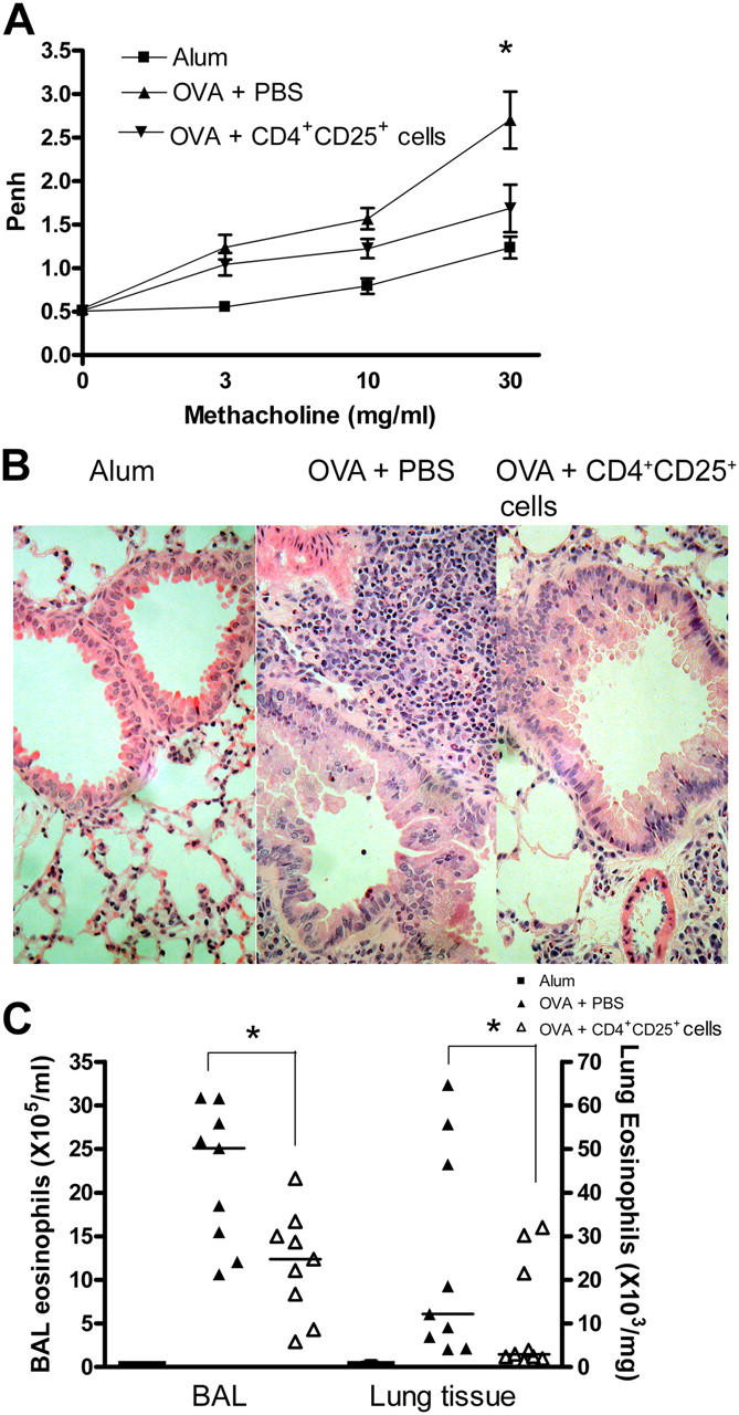
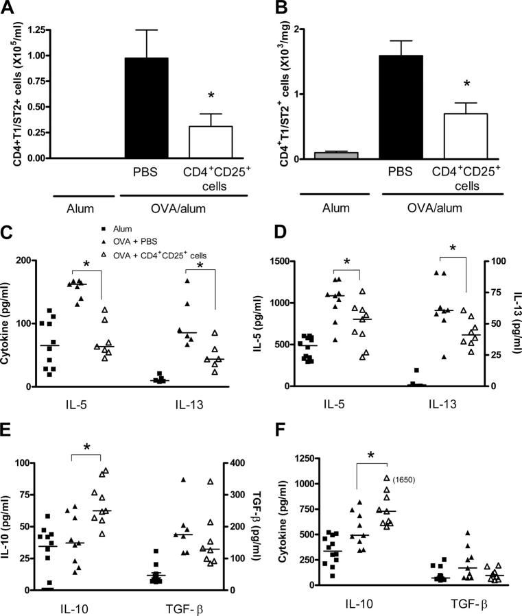
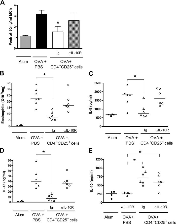
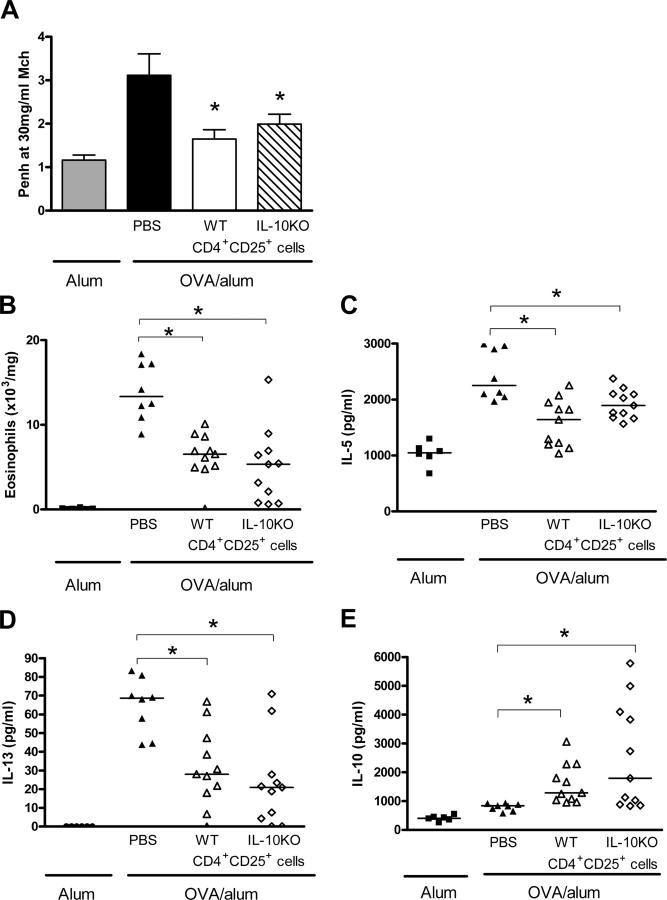
Deficient suppression of T cell responses to allergen by CD4+CD25+ regulatory T cells has been observed in patients with allergic disease. Our current experiments used a mouse model of airway inflammation to examine the suppressive activity of allergen-specific CD4+CD25+ T cells in vivo. Transfer of ovalbumin (OVA) peptide-specific CD4+CD25+ T cells to OVA-sensitized mice reduced airway hyperreactivity (AHR), recruitment of eosinophils, and T helper type 2 (Th2) cytokine expression in the lung after allergen challenge. This suppression was dependent on interleukin (IL) 10 because increased lung expression of IL-10 was detected after transfer of CD4+CD25+ T cells, and regulation was reversed by anti-IL-10R antibody. However, suppression of AHR, airway inflammation, and increased expression of IL-10 were still observed when CD4+CD25+ T cells from IL-10 gene-deficient mice were transferred. Intracellular cytokine staining confirmed that transfer of CD4+CD25+ T cells induced IL-10 expression in recipient CD4+ T cells, but no increase in IL-10 expression was detected in airway macrophages, dendritic cells, or B cells. These data suggest that CD4+CD25+ T cells can suppress the Th2 cell-driven response to allergen in vivo by an IL-10-dependent mechanism but that IL-10 production by the regulatory T cells themselves is not required for such suppression.


Figures

Figure 1.
Figure 1.
OVA-specific CD4+CD25+ regulatory T cells regulate allergen-induced airway inflammation in vivo. OVA-sensitized mice received either 5 × 10⁵ CD4+CD25+ cells or an equivalent volume of PBS and were challenged through the airways with OVA. (A) AHR was measured 24 h after the final OVA challenge using a Buxco system in which mice were exposed to increasing concentrations of methacholine. Values are expressed as means ± SEM (n = 9–12 mice/group from two separate experiments). *, P < 0.05 compared with OVA-sensitized mice that received PBS instead of CD4+CD25+ cells. (B) Lungs were fixed, sectioned, and stained with hemotoxylin and eosin. Representative sections are shown for each treatment group. BAL and lung tissue digest cells (C) were isolated as described in Materials and methods, and eosinophil numbers were determined by differential counts. Values are expressed as medians (n = 9–12 mice/group from two separate experiments). *, P < 0.05 compared with OVA-sensitized mice that received PBS instead of CD4+CD25+ cells.
Figure 2.
Figure 2.
Transfer of CD4+CD25+ regulatory T cells reduces Th2 cell responses in the lung after allergen challenge. BAL (A) and lung tissue digest cells (B) were isolated as described in Materials and methods. Th2 cell numbers were determined 24 h after the final OVA challenge by antibody staining and flow cytometric analysis, as described in Materials and methods. Th2 cells were defined as cells that were double-stained for CD4 and the Th2 cell–specific marker T1/ST2. Data are expressed as means ± SEM. IL-5 and IL-13 levels measured in BAL fluid (C) and lung homogenate supernatant (D) by ELISA. IL-10 and active TGF-β1 levels were measured in BAL fluid (E) and lung homogenate supernatant (F) by ELISA. Data are expressed as medians (n = 9–12 mice/group from two separate experiments). *, P < 0.05 compared with OVA-sensitized mice that received PBS instead of CD4+CD25+ cells.
Figure 3.
Figure 3.
Suppression of allergen-induced airway inflammation is IL-10 dependent. Mice were treated with anti–IL-10R antibody or control Ig during the allergen challenge phase of allergic inflammation. AHR (A) and lung eosinophilia (B) were quantified as described in Materials and methods. IL-5 (C), IL-13 (D), and IL-10 (E) levels were determined in lung homogenate supernatant by ELISA. Data are expressed as means ± SEM (A) or medians (B–E; n = 4–6 mice/group). *, P < 0.05 compared with OVA-sensitized mice that received PBS and control Ig.
Figure 4.
Figure 4.
Transfer of IL-10–deficient CD4+CD25+ regulatory T cells suppresses allergen-induced airway inflammation. Mice received either wild-type CD4+CD25+ regulatory T cells, IL-10–deficient CD4+CD25+ regulatory T cells, or an equivalent volume of PBS as a control. AHR (A) and lung eosinophilia (B) were determined as described in Materials and methods. IL-5 (C), IL-13 (D), and IL-10 (E) levels were measured in lung homogenate supernatant by ELISA. Data are expressed as means ± SEM (A) or medians (B-E; n = 6–11 mice/group from two separate experiments). *, P < 0.05 compared with OVA-sensitized mice that received PBS instead of CD4+CD25+ cells.
Figure 5.
Figure 5.
IL-10 is produced by CD4+ T cells during allergen-induced airway inflammation and is increased by transfer of CD4+CD25+ regulatory T cells. Lungs were digested with collagenase and DNase as described in Materials and methods. Digest cells were stimulated by PMA/Ionomycin in the presence of Brefeldin A for 6 h. Cells were phenotyped by staining for CD4, CD8, CD11b (macrophages), CD11c (dendritic cells), and B220 (B cells). Granulocytes were defined by forward and side scatter. (A) Data are expressed as median cell types producing IL-10 with interquartile range (n = 6–14 mice/group in three separate experiments). *, P < 0.05 compared with OVA-sensitized mice that received PBS instead of CD4+CD25+ cells. (B) Data are shown as representative FACS plots showing costaining of lung tissue digest cells with CD4 and IL-10. Percentages in the top right quadrants refer to median percentages of CD4 cells expressing IL-10 (n = 6–14 mice/group).

References

    1. Robinson, D.S., Q. Hamid, S. Ying, A. Tsicopoulos, J. Barkans, A.M. Bentley, C. Corrigan, S.R. Durham, and A.B. Kay. 1992. Predominant TH2-like bronchoalveolar T-lymphocyte population in atopic asthma. N. Engl. J. Med. 326:298–304. - PubMed
    1. Gavett, S.H., X. Chen, F. Finkelman, and M. Wills-Karp. 1994. Depletion of murine CD4+ T lymphocytes prevents antigen-induced airway hyperreactivity and pulmonary eosinophilia. Am. J. Respir. Cell Mol. Biol. 10:587–593. - PubMed
    1. Gonzalo, J.A., C.M. Lloyd, L. Kremer, E. Finger, A. Martinez, M.H. Siegelman, M. Cybulsky, and J.C. Gutierrez-Ramos. 1996. Eosinophil recruitment to the lung in a murine model of allergic inflammation. The role of T cells, chemokines, and adhesion receptors. J. Clin. Invest. 98:2332–2345. - PMC - PubMed
    1. Mosmann, T.R., and R.L. Coffman. 1989. TH1 and TH2 cells: different patterns of lymphokine secretion lead to different functional properties. Annu. Rev. Immunol. 7:145–173. - PubMed
    1. O'Garra, A., and P. Vieira. 2004. Regulatory T cells and mechanisms of immune system control. Nat. Med. 10:801–805. - PubMed

Publication types

MeSH terms