. 2005 Dec;11(12):1869-83.
doi: 10.1261/rna.2155905.

Biochemical analysis of the EJC reveals two new factors and a stable tetrameric protein core

Affiliations

Biochemical analysis of the EJC reveals two new factors and a stable tetrameric protein core

Thomas Ø Tange et al. RNA. 2005 Dec.

Abstract

The multiprotein exon junction complex (EJC) is deposited on mRNAs upstream of exon-exon junctions as a consequence of pre-mRNA splicing. In mammalian cells, this complex serves as a key modulator of spliced mRNA metabolism. To date, neither the complete composition nor the exact assembly pathway of the EJC has been entirely elucidated. Using in vitro splicing and a two-step chromatography procedure, we have purified the EJC and analyzed its components by mass spectrometry. In addition to finding most of the known EJC factors, we identified two novel EJC components, Acinus and SAP18. Heterokaryon analysis revealed that SAP18 is a shuttling protein whereas Acinus is restricted to the nucleus. In MS2 tethering assays Acinus stimulated gene expression at the RNA level, while MLN51, another EJC factor, stimulated mRNA translational efficiency. Using tandem affinity purification (TAP) of proteins overexpressed in HeLa cells, we demonstrated that Acinus binds directly to another EJC component, RNPS1, while stable association of SAP18 to form the trimeric apoptosis and splicing associated protein (ASAP) complex requires both Acinus and RNPS1. Using the same methodology, we further identified what appears to be the minimal stable EJC core, a heterotetrameric complex consisting of eIF4AIII, Magoh, Y14, and MLN51.


Figures

FIGURE 1.
FIGURE 1.
EJC purification. (A) Purification strategy. An AdML splicing substrate with six MS2 binding loops in the 3′ exon (AdML-6MS2) was spliced in nuclear extract supplemented with recombinant MS2-MBP and GST-Magoh:Y14. After splicing, the reaction was subject to RNase H cleavage (black lines beneath intron indicate the hybridization of the six DNA oligos used for the RNase H degradation of the unspliced pre-mRNA), gel filtration, and amylose affinity to purify the mRNP, which was then treated with RNase A followed by glutathione affinity to purify the GST-tagged EJC. (B) Coomassie-stained SDS polyacrylamide gel of purified proteins. Purified EJC proteins from 24 mL of in vitro splicing reactions containing either AdML-6MS2 pre-mRNA (lane 1) or intronless control AdML-6MS2mRNA (lane 2). The recombinant GST-Magoh:Y14 heterodimer used to supplement splicing reactions is also shown (lane 3).
FIGURE 2.
FIGURE 2.
Immuno-analysis of purified mRNP and EJC. (A) Western blot of the HeLa nuclear extract (lanes 1,2) used for the EJC purification and the amylose affinity purified mRNP (lane 3). The blot was first probed with antibody against MLN51 (upper panel), then stripped and reprobed with antibody against Upf3b (lower panel). (B) Western blot of HeLa nuclear extract (lanes 1,2) used for the EJC purification and the purified EJC (lane 3) with an antibody recognizing all three Acinus isoforms (positions as indicated).
FIGURE 3.
FIGURE 3.
Immunoprecipitation of in vitro splicing reactions. (A) Denaturing 15% gel showing AdML splicing products and immunoprecipitated RNA fragments. The positions of pre-mRNA, mRNA, debranched lariat, and the two fragments resulting from RNase H cleavage of the mRNA are indicated on the left. The round object depicts the deposited EJC. The line beneath the mRNA 5′ exon indicates the position of the DNA oligo used for RNase H digestion. Odd-numbered lanes are reactions with pre-mRNA and even-numbered lanes are reactions with unspliced control mRNA. (Lanes 1,2) Completed splicing reactions. (Lanes 3,4) RNase H digested reactions. (Lanes 5,6) Markers created by RNase H digestion of unspliced pre-mRNA and control mRNA for 10 min in NE supplemented with the DNA oligo (the center of the 12-nucleotide oligo is 49 nt upstream of the exon junction). (Lanes 722) Immunoprecipitated reactions using antibodies indicated above lanes. Lanes 14 contain half as much input RNA as lanes 722. (B) Denaturing 15% gel showing β-globin splicing products and immunoprecipitated RNA fragments. Odd- and even-numbered lanes contain reactions with 38- and 17-nt 5′ exon β-globin RNA, respectively. The migration positions of pre-mRNAs and mRNAs are indicated to the left. (Lanes 1,2) Unspliced pre-mRNAs. (Lanes 3,4) Completed splicing reactions. (Lanes 522) Immunoprecipitated reactions using the indicated antibodies. Lanes 3 and 4 contain half as much input RNA as lanes 522. The high background of β-globin pre-mRNA and lariat observed in all immunoprecipitations likely indicates a general affinity of the β-globin intron for protein G beads.
FIGURE 4.
FIGURE 4.
Heterokaryon analysis. SAP18 is a shuttling protein; AcinusL and AcinusS′ are nonshuttling proteins. (A) Localization of GFP-SAP18 (green) and nonshuttling control MS2-DEK (red) in a fused HeLa and a mouse NIH3T3 heterokaryon. The mouse nucleus is indicated by a white arrow in the DAPI-stained panel. (B,C) Localization of AcinusL-GFP (green) and AcinusS′-GFP (green) and the shuttling control MS2-hnRNP A1 (red) in a fused HeLa and a mouse NIH3T3 heterokaryon. The mouse nuclei are indicated by white arrows in the DAPI-stained panels. (D,E) Localization of Flag-AcinusL (red) and Flag-AcinusS′ (red) and the shuttling control eIF4AIII-GFP (green) in a fused HeLa and a mouse NIH3T3 heterokaryon. The mouse nuclei are indicated by white arrows in the DAPI-stained panels.
FIGURE 5.
FIGURE 5.
MS2 tethering assay. (A) Schematic of Renilla reporter with six MS2 binding loops at the 5′ end of the open reading frame and co-transfected firefly control. (B) RNase protection assay showing Renilla, firefly, and endogenous cyclophilin RNAs. Co-transfected MS2 fusion protein plasmids are indicated above. (C) Histogram depicting the combined results of three independent experiments. (White bars) Renilla RNA normalized to firefly RNA relative to cells expressing MS2 alone. (Gray bars) Renilla luciferase activity normalized to firefly activity relative to cells expressing MS2 alone. (Black bars) Gray bars/white bars = net effect on translational yield.
FIGURE 6.
FIGURE 6.
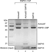
Silver-stained gel of purified complexes containing RNPS1-TAP, AcinusS′, and/or SAP18. HeLa cells were transfected with combinations of expression plasmids for the indicated proteins. The indicated TAP-tagged protein was purified along with bound factors by way of the Protein A (ProtA) and calmodulin binding protein (CBP) modules separated by a Tev proteinase cleavage site, which together constitute the TAP tag. (Lane 1) Co-expression of RNPS1-TAP and AcinusS′. (Lane 2) Co-expression of RNPS1-TAP, AcinusS′, and SAP18. (Lane 3) Co-expression of RNPS1-TAP and SAP18. Western blot shows the input levels of SAP18 in the three lysates prior to TAP purification.
FIGURE 7.
FIGURE 7.
Coomassie-stained gels of purified complexes containing Y14, Magoh, eIF4AIII, and/or MLN51. (A) Co-expression of Y14-TAP, Magoh, and eIF4AIII (lane 1); co-expression of Y14-TAP, Magoh, and Flag-MLN51 (lane 2); and co-expression of Y14-TAP, Magoh, eIF4AIII, and Flag-MLN51 (lane 3). Western blot panels show input levels of Flag-MLN51 and eIF4AIII in all lysates prior to TAP purification. (B) Proteins purified upon co-expression of Y14-TevProtA, Magoh, eIF4AIII-CBP, and Flag-MLN51. Western blot panels show input levels of Flag-MLN51, eIF4AIII-CBP, eIF4AIII (endogenous), and Y14-TevProtA (detected by α-eIF4AIII via the ProtA tag) in the lysate prior to TAP purification.
FIGURE 8.
FIGURE 8.
Three spheres of EJC factors. The minimal EJC core likely consists of a tetrameric complex containing eIF4AIII, MLN51, Magoh, and Y14 (this study), with eIF4AIII providing direct contact to the mRNA (Shibuya et al. 2004). All factors in this core are shuttling proteins and most likely follow the mRNA to the cytoplasm (Kataoka et al. 2000, 2001; Le Hir et al. 2001a,b; Macchi et al. 2003; Degot et al. 2004; Palacios et al. 2004; Shibuya et al. 2004). Proteins in the outer shell were all found by mass spectrometry of the in vitro-derived EJC (this study). RNPS1, Acinus, and SAP18 can stably associate (Schwerk et al. 2003) and may bind the EJC core as a trimeric complex. However, RNPS1 may also bind alone, e.g., via interactions with Pinin (Sakashita et al. 2004). SAP18, RNPS1, and Aly/REF are shuttling proteins (this study; Zhou et al. 2000; Lykke-Andersen et al. 2001), whereas Acinus and Pinin are nuclear restricted (this study; Li et al. 2003). Transiently interacting factors are proteins not identified in the in vitro-derived EJC, but which likely interact dynamically with either the EJC core or outer sphere proteins (see text).

Similar articles

Cited by

References

    1. Ballut, L., Marchadier, B., Baguet, A., Tomasetto, C., Seraphin, B., and Le Hir, H. 2005. The exon junction core complex is locked onto RNA by inhibition of EIF4AIII ATPase activity. Nat. Struct. Mol. Biol. 12: 861–869. - PubMed
    1. Chan, C.C., Dostie, J., Diem, M.D., Feng, W., Mann, M., Rappsilber, J., and Dreyfuss, G. 2004. eIF4A3 is a novel component of the exon junction complex. RNA 10: 200–209. - PMC - PubMed
    1. Cheng, S.Y. and Bishop, J.M. 2002. Suppressor of Fused represses Gli-mediated transcription by recruiting the SAP18-mSin3 corepressor complex. Proc. Natl. Acad. Sci. 99: 5442–5447. - PMC - PubMed
    1. Conti, E. and Izaurralde, E. 2005. Nonsense-mediated mRNA decay: Molecular insights and mechanistic variations across species. Curr. Opin. Cell Biol. 17: 316–325. - PubMed
    1. Degot, S., Le Hir, H., Alpy, F., Kedinger, V., Stoll, I., Wendling, C., Seraphin, B., Rio, M.C., and Tomasetto, C. 2004. Association of the breast cancer protein MLN51 with the exon junction complex via its speckle localizer and RNA binding module. J. Biol. Chem. 279: 33702–33715. - PubMed

Publication types

MeSH terms