. 2005 Dec;167(6):1575-86.
doi: 10.1016/S0002-9440(10)61242-4.

Biomimetic delivery of keratinocyte growth factor upon cellular demand for accelerated wound healing in vitro and in vivo

Affiliations

Biomimetic delivery of keratinocyte growth factor upon cellular demand for accelerated wound healing in vitro and in vivo

David J Geer et al. Am J Pathol. 2005 Dec.

Abstract

Exogenous keratinocyte growth factor (KGF) significantly enhances wound healing, but its use is hampered by a short biological half-life and lack of tissue selectivity. We used a biomimetic approach to achieve cell-controlled delivery of KGF by covalently attaching a fluorescent matrix-binding peptide that contained two domains: one recognized by factor XIII and the other by plasmin. Modified KGF was incorporated into the fibrin matrix at high concentration in a factor XIII-dependent manner. Cell-mediated activation of plasminogen to plasmin degraded the fibrin matrix and cleaved the peptides, releasing active KGF to the local microenvironment and enhancing epithelial cell proliferation and migration. To demonstrate in vivo effectiveness, we used a hybrid model of wound healing that involved transplanting human bioengineered skin onto athymic mice. At 6 weeks after grafting, the transplanted tissues underwent full thickness wounding and treatment with fibrin gels containing bound KGF. In contrast to topical KGF, fibrin-bound KGF persisted in the wounds for several days and was released gradually, resulting in significantly enhanced wound closure. A fibrinolytic inhibitor prevented this healing, indicating the requirement for cell-mediated fibrin degradation to release KGF. In conclusion, this biomimetic approach of localized, cell-controlled delivery of growth factors may accelerate healing of large full-thickness wounds and chronic wounds that are notoriously difficult to heal.


Figures
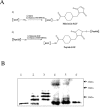
Figure 1
Figure 1
Functional analysis of KGF for binding to fibrin gels and Western analysis of products. A: KGF was functionalized with 1.7-kd peptides by a two-step reaction. In the first reaction (1), KGF was derivatized with a heterobifunctional linker molecule (SMCC) at a 10:1 (SMCC:KGF) molecular ratio, yielding a reactive maleimido-KGF. After purification, a second reaction (2) involved the peptide conjugation to maleimide-derivatized KGF to form a stable thioester bond yielding PN-KGF (N = molecular ratio of peptides to KGF molecules reacted). B: Western analysis was used to examine the KGF protein (18.9 kd) after modification and release from fibrin gels through the action of plasmin. Lane 1, stock KGF was used as control; lane 2, P2-KGF; lane 3, P5-KGF; lane 4, PL-P2-KGF; lane 5, PL-P5-KGF, after conjugation to fibrin, the gels were washed extensively and degraded with 0.25 U/ml of plasmin at 37°C for 3 hours; lane 6, soluble P5-KGF (not conjugated to fibrin) was treated with 0.25 U/ml plasmin at 37°C for 3 hours.
Figure 2
Figure 2
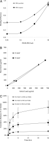
Fibrin hydrogels have high binding capacity and the binding efficiency is dose-dependent on factor XIII. A: P-KGF was added to fibrin gels in the presence of factor XIII at the indicated concentrations (0.01 to 10 PEU/ml). Impure and purified fibrinogen preparations were used for comparison. Gels were thoroughly washed in buffer and then degraded with plasmin at 37°C for 3 hours. Total fluorescence measurements of the gel components before gel formation and after plasmin degradation were used to calculate the binding efficiency. B: The amount of bound P-KGF in fibrin matrix increases linearly with the amount added up to 500 μg/ml. C: Kinetics of KGF release from Fb-KGF and Fb-P-KGF formulations that were polymerized in the presence of 0 or 10 PEU/ml of factor XIII. Values are the mean ± SD of triplicate samples in a representative experiment (n = 3).
Figure 3
Figure 3
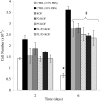
Modified KGF promotes proliferation of epithelial cells. KGF samples (stock KGF, P2-KGF, P5-KGF, PL-P2-KGF, and PL-P5-KGF) were added to the wells of a 96-well plate at 500 ng/ml and 4Mbr-5 lung epithelial cells (10,000 per well) were then seeded in low-serum media (CTRL, 2.5% FBS). For comparison, cells were grown in regular, high-serum media (CTRL, 10% FBS) with 30 ng/ml of epidermal growth factor. At 2 and 6 days after seeding, plates were washed once and frozen for Hoechst DNA quantitation. Total DNA was measured at the indicated times using a fluorescence microplate reader. The number of cells was determined from fluorescence measurements using a standard curve. Values are the mean ± SD of triplicate samples in a representative experiment (n = 3). An asterisk indicates a significant decrease (P = 0.0002) in growth at 6 days as compared to 2 days. A double dagger indicates a significant increase (P < 0.001) in growth for all samples with unmodified and modified KGF as compared to CTRL (2.5%) at 6 days.
Figure 4
Figure 4
Cell-mediated release of KGF from fibrin gels promotes wound closure in vitro. Lung epithelial 4MBr-5 cells were grown on collagen-coated plates. At confluence the monolayers were wounded and overlaid with 300 μl per well of Fb or Fb-P-KGF (150 ng of P-KGF) in the presence of 0, 10, or 100 kIU/ml aprotinin. The solutions polymerized within 10 to 20 seconds after addition in the wells. The rate of healing was determined by measuring the denuded area in the wound at various times after wounding and dividing by the initial area. All values are the mean ± SD of triplicate samples in a representative experiment (n = 2).
Figure 5
Figure 5
Fluorescence imaging of wounds in vivo. Skin equivalents were grafted to athymic mice and allowed to take for 6 weeks before subjected to a full-thickness 4.0-mm excisional wound. An inverted fluorescence microscope was used to image the Fb-P-KGF or P-KGF in the wound (W) and periphery (P). Wounds were treated with Fb-P-KGF and imaged at 2 days (A) and 7 days (B) after wounding. Other wounds were treated by topical application of P-KGF (without fibrin gel) and imaged at 1 day (C). D: Tissues treated with Fb-P-KGF were excised at 8 days after wounding and processed for histology. Overlaid fluorescent and bright-field images of the wound show migrating cells as they degrade the fibrin matrix (green). Arrows represent wound edges and an asterisk denotes tip of migrating cells. Original magnifications: ×4 (A–C); ×10 (D).
Figure 6
Figure 6
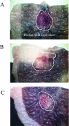
Macroscopic evaluation of wounded tissues. Skin equivalents were grafted to athymic mice and allowed to take for 6 weeks before subjected to a full-thickness 4.0-mm excisional wound. At 8 days after wounding, the excised tissues were placed on a stereomicroscope and the wounds were photographed with a digital camera. Wounds were treated with Fb (A), Fb-KGF (B), or Fb-P-KGF (C). The grafted human skin equivalent is dark because the cells were harvested from a dark-skin donor. Solid lines represent the original wound edges. Dotted lines indicate portion of the tissue that did not heal.
Figure 7
Figure 7
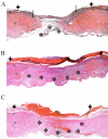
Histological evaluation of wounded tissues. Skin equivalents were grafted to athymic mice and allowed to take for 6 weeks before subjected to a full-thickness 4-mm excisional wound. At 8 days after wounding, tissues were excised and processed for H&E staining. Wounds treated with Fb (A), Fb-KGF (B), or Fb-P-KGF (C). A montage of images was created to facilitate visualization of the whole wound and evaluation of healing. Short arrows demarcate original wound edges, long arrows represent the tip of the migrating epithelium, and asterisks denote granulation tissue. Morphological features are indicated by E, epidermis; D, dermis; and Fb, fibrin hydrogel. Original magnifications, ×4.
Figure 8
Figure 8
Cell-mediated release of KGF from fibrin gels accelerates wound healing in vivo. Skin equivalents were grafted to athymic mice and allowed to take for 6 weeks before subjected to a full-thickness 4-mm excisional wound. The wounds were immediately treated with Fb (n = 7), Fb-KGF (n = 8), or Fb-P-KGF (n = 6). At 8 days after wounding, tissues were excised and processed for H&E staining. A: Re-epithelialization of the tissues was assessed by measuring the length of the migratory epithelium in the wound and dividing by the total wound size (% healing). B: Aprotinin was added at 100 or 1000 kIU/ml in Fb-KGF or Fb-P-KGF (n = 2). Results were averaged from histological measurements in two independent experiments. An asterisk indicates significant difference between Fb-KGF (P = 0.00047) or Fb-P-KGF (P = 0.013) as compared to Fb controls.

References

    1. Coulombe PA. Towards a molecular definition of keratinocyte activation after acute injury to stratified epithelia. Biochem Biophys Res Commun. 1997;236:231–238. - PubMed
    1. Collen D. Ham-Wasserman lecture: role of the plasminogen system in fibrin-homeostasis and tissue remodeling. Hematology (Am Soc Hematol Educ Program) 2001:1–9. - PubMed
    1. Aaronson SA, Bottaro DP, Miki T, Ron D, Finch PW, Fleming TP, Ahn J, Taylor WG, Rubin JS. Keratinocyte growth factor. A fibroblast growth factor family member with unusual target cell specificity. Ann NY Acad Sci. 1991;638:62–77. - PubMed
    1. Werner S. Keratinocyte growth factor: a unique player in epithelial repair processes. Cytokine Growth Factor Rev. 1998;9:153–165. - PubMed
    1. Rubin JS, Bottaro DP, Chedid M, Miki T, Ron D, Cheon G, Taylor WG, Fortney E, Sakata H, Finch PW, LaRochelle WJ. Keratinocyte growth factor. Cell Biol Int. 1995;19:399–411. - PubMed

Publication types

LinkOut - more resources