. 2005 Dec;25(24):10833-41.
doi: 10.1128/MCB.25.24.10833-10841.2005.

The essential WD40 protein Cia1 is involved in a late step of cytosolic and nuclear iron-sulfur protein assembly

Affiliations

The essential WD40 protein Cia1 is involved in a late step of cytosolic and nuclear iron-sulfur protein assembly

Janneke Balk et al. Mol Cell Biol. 2005 Dec.

Abstract

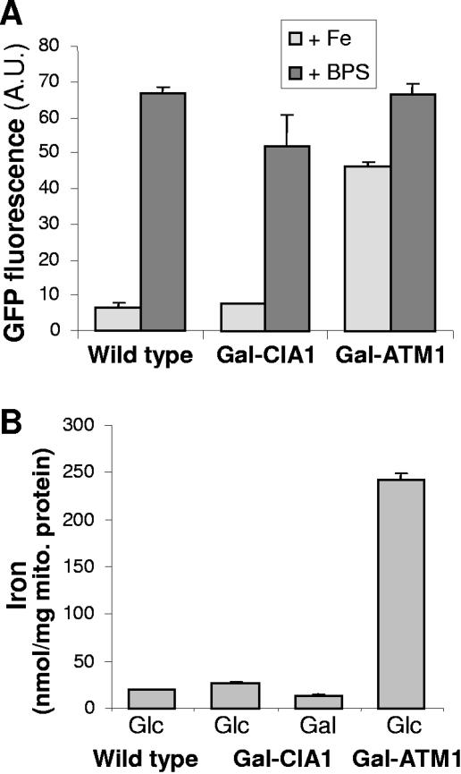
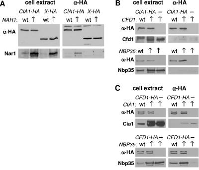
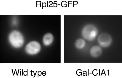
The assembly of cytosolic and nuclear iron-sulfur (Fe/S) proteins in yeast is dependent on the iron-sulfur cluster assembly and export machineries in mitochondria and three recently identified extramitochondrial proteins, the P-loop NTPases Cfd1 and Nbp35 and the hydrogenase-like Nar1. However, the molecular mechanism of Fe/S protein assembly in the cytosol is far from being understood, and more components are anticipated to take part in this process. Here, we have identified and functionally characterized a novel WD40 repeat protein, designated Cia1, as an essential component required for Fe/S cluster assembly in vivo on cytosolic and nuclear, but not mitochondrial, Fe/S proteins. Surprisingly, Nbp35 and Nar1, themselves Fe/S proteins, could assemble their Fe/S clusters in the absence of Cia1, demonstrating that these components act before Cia1. Consequently, Cia1 is involved in a late step of Fe/S cluster incorporation into target proteins. Coimmunoprecipitation assays demonstrated a specific interaction between Cia1 and Nar1. In contrast to the mostly cytosolic Nar1, Cia1 is preferentially localized to the nucleus, suggesting an additional function of Cia1. Taken together, our results indicate that Cia1 is a new member of the cytosolic Fe/S protein assembly (CIA) machinery participating in a step after Nbp35 and Nar1.


Figures

FIG. 1.
FIG. 1.
Schematic representation of CFD1 and CIA1 genes in S. cerevisiae and S. pombe. A protein-coding sequence highly similar to S. cerevisiae CFD1 is found in S. pombe as a fusion gene with a C-terminal WD40 repeat domain (systematic name, SPAC806.02).
FIG. 2.
FIG. 2.
The essential Cia1 is a soluble protein in the nucleus and cytosol. (A) Growth of Cia1-depleted cells. Gal-CIA1 cells carrying a galactose-regulatable (Gal) CIA1 gene were transformed with vector p416 without or with a CIA1-HA fusion gene. Cells were grown for 2 × 2 days at 30°C on agar plates containing rich media supplemented with galactose (YPGal) or glucose (YPD). Tenfold serial dilutions are shown. (B) Subcellular fractionation and immunoblot analysis. Wild-type cells were disrupted by removal of the cell wall and homogenization with a Douncer in 0.6 M sorbitol, 20 mM HEPES-KOH, pH 7.4, 1 mM dithiothreitol, and protease inhibitors, followed by differential centrifugation. After discarding a low-speed pellet containing unbroken cells, nuclei, and debris, the cell extract (CE) was centrifuged at 10,000 × g for 10 min to separate soluble proteins (S10) from a fraction enriched in organelles (P10). The S10 fraction was further centrifuged at 100,000 × g for 30 min to remove membranes (P100) from soluble proteins (S100). Nuclei (Nuc) were purified separately using a Ficoll density gradient (1). Equal amounts of protein (20 μg/per lane) were separated by sodium dodecyl sulfate-polyacrylamide gel electrophoresis, blotted, and immunostained for Cia1 or marker proteins of the nucleus (DNA polymerase-associated Pol30), mitochondria (Mge1), endoplasmic reticulum (translocon subunit Sec61), and the cytosol (Leu1). (C) In situ localization of Cia1. Wild-type cells were transformed with the high-copy-number expression vector p426 containing CIA1 fused to the HA tag sequence. Log-phase cells were fixed with 2.4% (wt/vol) formaldehyde, permeabilized, and labeled with monoclonal anti-HA (α-HA) antibodies, followed by fluorophore-conjugated secondary antibodies. DNA was counterstained with DAPI (4′,6′-diamidino-2-phenylindole) to show the positions of the nucleus and mitochondria.
FIG. 3.
FIG. 3.
Cia1 is required for the assembly of a subset of cytosolic and nuclear Fe/S proteins. Gal-CIA1 cells were grown in minimal medium supplemented with galactose (Gal) or glucose (Glc) for various lengths of time to induce or suppress, respectively, expression of CIA1. (A) Enzyme activities of alcohol dehydrogenase (ADH), isopropylmalate isomerase (Leu1), and sulfite reductase (SiR) in cell extracts are presented relative to the values obtained for wild-type cells grown with galactose. The protein levels of Cia1 and Leu1 were visualized by immunoblot analysis (lower panels). (B) 55Fe incorporation into Leu1. Gal-CIA1 cells were labeled with 55Fe, and a cell lysate was prepared, from which Leu1 was immunoprecipitated with specific antibodies. Coprecipitated 55Fe was quantified by scintillation counting. Immunoprecipitation with preimmune serum (PIS) was used as a control. The amounts of Leu1 protein were quantified by immunoblot analysis using chemiluminescence and densitometry of subsaturated film exposures. (C) 55Fe incorporation into several cytosolic and nuclear Fe/S proteins. Data acquired as in panel B are expressed as the ratio of glucose-grown (40 h) to galactose-grown cells, corrected for background, for each of the reporter proteins. Rli1-HA, Ntg2-HA, Nbp35-TAP, and Nar1 were expressed from high-copy-number vectors (p426) and immunoprecipitated with anti-HA, immnunoglobulin G (IgG), and anti-Nar1 antibodies, respectively. Cells not expressing these constructs were used to determine the background levels of immunoprecipitated 55Fe. The panels on the right show examples of the immunoblot analysis for each of the indicated proteins.
FIG. 4.
FIG. 4.
Cia1 is not required for mitochondrial Fe/S protein assembly. (A) Enzyme activities of aconitase and succinate dehydrogenase (SDH) in wild-type (WT) and Gal-CIA1 cells after growth in minimal medium plus glucose for 40 h. Protein levels of aconitase (Aco1) and succinate dehydrogenase subunit 2 (Sdh2) were visualized by immunoblot analysis (inset), using amounts of mitochondrial protein equivalent to those used for the enzyme assays. (B) 55Fe incorporation into the Fe/S proteins Bio2 and Yah1-HA. Gal-CIA1 cells were grown in iron-poor minimal medium supplemented with galactose (Gal) or glucose (Glc) to induce or suppress, respectively, expression of CIA1. Bio2 and HA-tagged Yah1 were overproduced using the high-copy-number vector p426. Cells were labeled with 55Fe, and a cell lysate was prepared, from which Bio2 and Yah1 were immunoprecipitated with anti-Bio2- or anti-HA-specific antibodies, respectively. Coprecipitated 55Fe was quantified by scintillation counting. The amounts of 55Fe immunoprecipitated from cell extracts that did not contain overproduced Bio2 or Yah1-HA were taken as background (Bg). The levels of the indicated proteins were analyzed by immunoblot analysis.
FIG. 5.
FIG. 5.
Normal cellular and mitochondrial iron homeostasis in the absence of Cia1. (A) Wild-type, Gal-CIA1, and Gal-ATM1 cells carrying plasmid p415-FET3-GFP were grown in glucose-containing minimal medium for 24 h, diluted to an optical density at 600 nm of 0.1, and supplemented with either 0.1 mM FeCl3 (Fe) or 0.1 mM of the iron chelator bathophenanthroline sulfonate (BPS). Growth was continued for a further 6 h. The transcriptional activity of the FET3 promoter was determined by recording the fluorescence emission of the cell suspension at 513 nm (excitation at 480 nm). The signal of cells lacking plasmid p415-FET3-GFP was subtracted. A.U., arbitrary units. (B) Wild-type, Gal-CIA1, and Gal-ATM1 cells were grown in minimal medium with either galactose (Gal) or glucose (Glc). Mitochondria were purified by differential centrifugation, and the total iron content was measured using a colorimetric assay based on the iron chelator ferene.
FIG. 6.
FIG. 6.
Cia1 undergoes a protein interaction with Nar1. Cells producing the HA-tagged proteins as indicated were grown in minimal medium and harvested, and a cell extract was prepared. HA-agarose was added for 1 h of incubation at 4°C and washed several times with buffer. Both the cell extract and the washed HA-agarose were subjected to protein blot analysis to detect proteins of interest. (A) Coimmunoprecipitation of HA-tagged Cia1 with Nar1 in extracts from cells with wild-type (wt) or overproduced (↑) levels of Nar1. Another HA-tagged protein was used as a control (X-HA, where X represents Ymr134w). The indicated proteins were detected by protein blot analysis in total cell extracts (left panels) and after immunoprecipitation (anti-HA [α-HA], right panels). (B and C) As in panel A, using cells overproducing Cfd1, Nbp35, or Cia1 as indicated. Cia1-HA (B) or Cfd1-HA (C) was immunoprecipitated using anti-HA antibodies before protein blot analyses.
FIG. 7.
FIG. 7.
Cia1 is required for export of the large ribosomal subunit from the nucleus. Wild-type and Gal-CIA1 cells expressing an RPL25-GFP fusion gene were cultured in minimal medium supplemented with glucose for 40 h. The cellular distribution of GFP was monitored by fluorescence microscopy. The spot accumulating GFP in the Gal-CIA1 cells is the nucleus, as judged by phase-contrast microscopy and DAPI (4′,6′-diamidino-2-phenylindole) staining (not shown; see reference 16).

References

    1. Aris, J. P., and G. Blobel. 1991. Isolation of yeast nuclei. Methods Enzymol. 194:735-749. - PubMed
    1. Babcock, M., D. de Silva, R. Oaks, S. Davis-Kaplan, S. Jiralerspong, L. Montermini, M. Pandolfo, and J. Kaplan. 1997. Regulation of mitochondrial iron accumulation by Yfh1p, a putative homolog of frataxin. Science 276:1709-1712. - PubMed
    1. Balk, J., A. J. Pierik, D. J. Aguilar Netz, U. Mühlenhoff, and R. Lill. 2004. The hydrogenase-like Nar1p is essential for maturation of cytosolic and nuclear iron-sulphur proteins. EMBO J. 23:2105-2115. - PMC - PubMed
    1. Balk, J., A. J. Pierik, D. J. Aguilar Netz, U. Mühlenhoff, and R. Lill. 2005. Nar1p, a conserved eukaryotic protein with similarity to Fe-only hydrogenases, functions in cytosolic iron-sulphur protein biogenesis. Biochem. Soc. Trans. 33:86-89. - PubMed
    1. Ghaemmaghami, S., W.-K. Huh, K. Bower, R. W. Howson, A. Belle, N. Dephoure, E. K. O'Shea, and J. S. Weissman. 2003. Global analysis of protein expression in yeast. Nature 425:737-741. - PubMed

Publication types

MeSH terms