. 2005 Dec;25(24):10916-29.
doi: 10.1128/MCB.25.24.10916-10929.2005.

Antagonistic effects of Grg6 and Groucho/TLE on the transcription repression activity of brain factor 1/FoxG1 and cortical neuron differentiation

Affiliations

Antagonistic effects of Grg6 and Groucho/TLE on the transcription repression activity of brain factor 1/FoxG1 and cortical neuron differentiation

Nathalie Marçal et al. Mol Cell Biol. 2005 Dec.

Abstract

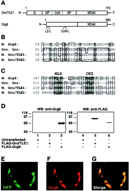
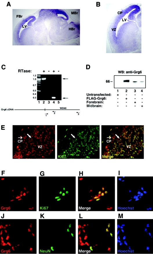
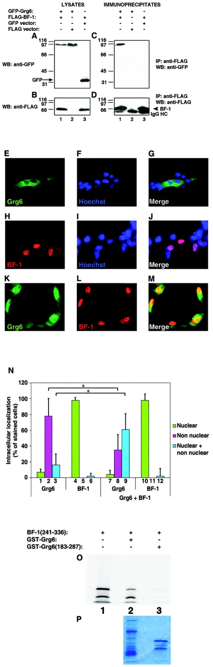
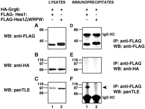
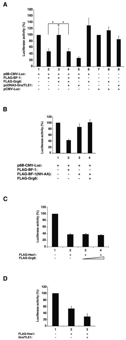
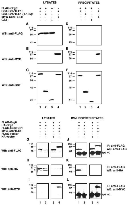
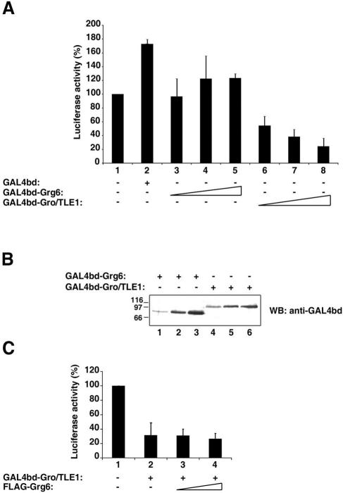
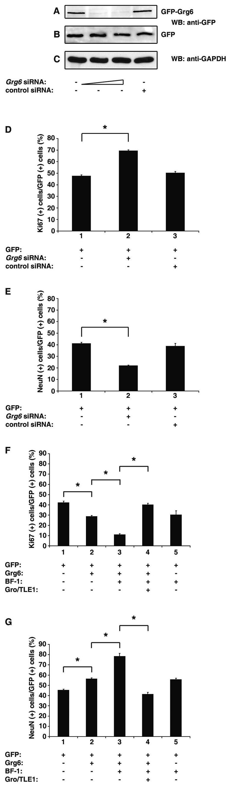
Groucho (Gro)/TLE transcriptional corepressors are involved in a variety of developmental mechanisms, including neuronal differentiation. They contain a conserved C-terminal WD40 repeat domain that mediates interactions with several DNA-binding proteins. In particular, Gro/TLE1 interacts with forkhead transcription factor brain factor 1 (BF-1; also termed FoxG1). BF-1 is an essential regulator of neuronal differentiation during cerebral cortex development and represses transcription together with Gro/TLE1. Gro/TLE-related gene product 6 (Grg6) shares with Gro/TLEs a conserved WD40 repeat domain but is more distantly related at its N-terminal half. We demonstrate that Grg6 is expressed in cortical neural progenitor cells and interacts with BF-1. In contrast to Gro/TLE1, however, Grg6 does not promote, but rather suppresses, BF-1-mediated transcriptional repression. Consistent with these observations, Grg6 interferes with the binding of Gro/TLE1 to BF-1 and does not repress transcription when targeted to DNA. Moreover, coexpression of Grg6 and BF-1 in cortical progenitor cells leads to a decrease in the number of proliferating cells and increased neuronal differentiation. Conversely, Grg6 knockdown by RNA interference causes decreased neurogenesis. These results identify a new role for Grg6 in cortical neuron development and establish a functional link between Grg6 and BF-1.


Figures

FIG. 1.
FIG. 1.
Characterization of anti-Grg6 antibody. (A) Top, schematic representation of the domain structure of Gro/TLE1 (37); bottom, Grg6 displays the highest relatedness to Gro/TLEs at the level of the C-terminal WDR domain, with similarity within the N-terminal half limited only to a potential leucine zipper-like motif (LZ-L) and a CcN domain-like region (CcN-L). (B) Comparison of the putative LZ-L motif of mouse (M.) Grg6 and the first LZ-L motif within the Q domain of Drosophila (Dro.) Gro and mouse Gro/TLE1 and Gro/TLE2. Hydrophobic residues considered to form the core of this motif (36) are shaded and in bold. Identical amino acids and conservative substitutions are boxed. (C) Comparison of the CcN motif of Gro/TLE proteins and the CcN-L motif of Grg6. The nuclear localization sequence (NLS) and protein kinase CK2 phosphorylation site (CK2) (27) are shaded and in bold. Identical amino acids and conservative substitutions are boxed. (D) Western blotting analysis. HEK293 cells were either not transfected (lanes 1 and 4) or transfected with FLAG-Gro/TLE1 (lanes 2 and 5) or FLAG-Grg6 (lanes 3 and 6), followed by Western blotting (WB) with anti-Grg6 (lanes 1 to 3) or anti-FLAG (lanes 4 to 6) antibodies (Ab). Here and in succeeding figures, the positions and sizes of standards are indicated in kilodaltons. (E to G) COS7 cells were transfected with GFP-Grg6, fixed, and subjected to double-labeling analysis of GFP expression (E) and anti-Grg6 immunoreactivity (F). (G) Combined GFP and Grg6 staining.
FIG.2.
FIG.2.
Grg6 expression in the telencephalon. (A and B) In situ hybridization studies. Sagittal sections through the brains of E14.5 mouse embryos were analyzed with an antisense Grg6 riboprobe. Hybridization signals are observed in the forebrain (FBr), dorsal midbrain (Mbr), and caudal hindbrain (HBr); LV, lateral ventricle. No hybridization was detected with control sense riboprobes (not shown). (B) Higher-magnification view of the telencephalon showing robust Grg6 expression in both the ventricular zone (VZ) and the cortical plate (CP). (C) RT-PCR analysis. Each RNA was incubated with (lanes 2 and 4) or without (lanes 3 and 5) reverse transcriptase (RTase). The ensuing cDNA mixture was subjected to PCR with either primers 1 and 3 (lanes 2 and 3) or primers 1 and 2 (lanes 4 and 5). Lane 1 was loaded with the indicated DNA size standards (sizes are in kilobases). A schematic of the Grg6 cDNA is shown, indicating the location of the region encoding the WDR domain and the positions of the oligonucleotide primers. (D) Western blotting analysis. Lysates from HEK293 cells left untransfected (lane 1) or transfected with FLAG-Grg6 (lane 2) were subjected to SDS-PAGE together with lysates from forebrain (lane 3) or midbrain (lane 4) tissue dissected from E15.5 mouse embryos, followed by Western blotting (WB) with affinity-purified anti-Grg6 antibody. (E) Double-labeling immunohistochemical analysis of the dorsal telencephalon from E14.5 mouse embryos with antibodies against Grg6 (left) and Ki67 (middle). Combined Grg6-Ki67 staining is shown on the right; the large arrow points to an example of a double-labeled cell, while the small arrow points to cells positive for Grg6 but not Ki67 expression. Mitotic cells of the surface ectoderm are also visible in the top left corner. (F to M) Primary cultures of E13.5 mouse embryonic cortical progenitor cells grown for 4 days in vitro were fixed and subjected to double-labeling analysis of the expression of Grg6 (F and J), Ki67 (G), and NeuN (K). Combined Grg6-Ki67 (H) or Grg6-NeuN (L) staining is shown. Hoechst staining was used to visualize nuclei (I and M).
FIG.3.
FIG.3.
Interaction of Grg6 and BF-1. (A to D) Coimmunoprecipitation studies. HEK293 cells were transfected with FLAG-BF-1 in the absence or presence of GFP-Grg6 or GFP alone, as indicated. Each cell lysate was subjected to immunoprecipitation (IP) with anti-FLAG antibody (C and D), followed by analysis of the immunoprecipitated material, together with 1/10 of each input lysate (A and B), by Western blotting (WB) with anti-GFP (A and C) or anti-FLAG (B and D) antibodies. In panel A, the arrow points to the position of migration of GFP alone. In panel D, the arrowhead points to the position of migration of BF-1. Here and in succeeding figures, IgG HC indicates the immunoglobulin G heavy chain. (E to M) Immunocytochemical analysis. COS7 cells were transfected with GFP-Grg6 (E to G), FLAG-BF-1 (H to J), or the two proteins together (K to M), followed by double-labeling analysis of the expression of GFP-Grg6 (E and K) or BF-1 (H and L). (M) Combined GFP-Grg6/BF-1 staining. Hoechst staining is shown alone (F and I) or in combination with GFP-Grg6 (G) or BF-1 (J). (N) Separate immunocytochemical experiments (n = 7) were used to quantitate the intracellular distribution of Grg6 in the absence (bars 1 to 3) or presence (bars 7 to 9) of BF-1. The localization of BF-1 in the absence (bars 4 to 6) or presence (bars 10 to 12) of Grg6 is also shown. Results are depicted as the mean ± the standard deviation. *, P < 0.01 by analysis of variance. (O and P) In vitro interaction of Grg6 and BF-1. Amino acids 241 to 336 of BF-1 were in vitro translated as a fusion protein with GAL4bd and incubated with ∼2.0 μg of GST-Grg6 (lane 2) or GST-Grg6(183-287) (lane 3) purified from bacteria. (O) Precipitates recovered with glutathione-Sepharose beads were subjected to SDS-PAGE and autoradiography together with 10% of the amount of the in vitro-translated protein used in the incubation mixture (lane 1). GAL4-BF-1(241-336) consistently migrated as a doublet. (P) Coomassie blue staining of the GST fusion proteins used in these assays.
FIG. 4.
FIG. 4.
Interaction of Hes1 with Gro/TLE but not Grg6. HEK293 cells were cotransfected with HA-Grg6 and either FLAG-Hes1 (lane 1) or FLAG-Hes1ΔWRPW (lane 2). Each cell lysate was subjected to immunoprecipitation (IP) with anti-FLAG antibody (D to F), followed by analysis of the immunoprecipitates, together with 1/10 of each input lysate collected prior to immunoprecipitation (A to C), by Western blotting (WB) with anti-FLAG (A and D), anti-HA (B and E), or panTLE (C and F) antibodies. In panel F, the arrowhead points to the position of migration of endogenous Gro/TLE proteins.
FIG. 5.
FIG. 5.
Effect of Grg6 on BF-1-mediated transcriptional repression. (A and B) HEK293 cells were transfected with either the reporter construct p6B-CMV-Luc (A, bars1 to 6; B, bars 1 to 4) or the control plasmid pCMV-Luc (A, bars 7 to 9) in the absence or presence of the indicated combinations of expression vectors. (C) HEK293 cells were transfected with the reporter construct p6N-βactin-Luc in the absence (bar 1) or presence (bar 2) of FLAG-Hes1 and either 25 ng/transfection (bar 3) or 50 ng/transfection (bar 4) of FLAG-Grg6. (D) Cells were transfected as in panel C, except that a Gro/TLE1 expression plasmid (50 ng/transfection) was used instead of Grg6. In all panels, basal luciferase activity in the absence of effector plasmids was considered 100% and values in the presence of effector plasmids are expressed as the mean ± the standard deviation of at least three independent experiments performed in duplicate. (A) *, P < 0.05 by analysis of variance; the difference between bars 1 and 6 was not statistically significant.
FIG. 6.
FIG. 6.
Decreased interaction of BF-1 with Gro/TLE in the presence of Grg6. (A to F) HEK293 cells were transfected with the indicated combinations of expression vectors, followed by immunoprecipitation (IP) of endogenous Gro/TLE1 proteins with anti-Gro/TLE1 antibodies. The immunoprecipitates (D to F), together with 1/10 of each input lysate collected prior to immunoprecipitation (A to C), were subjected to Western blotting (WB) with either anti-FLAG (A and D), anti-Grg6 (B and E), or panTLE (C and F) antibodies. (G to J) HEK293 cells were transfected with the indicated combinations of expression vectors, followed by immunoprecipitation of FLAG-BF-1. The immunoprecipitates (I and J), together with 1/10 of each input lysate collected prior to immunoprecipitation (G and H), were subjected to Western blotting with either anti-GFP (G and I) or anti-FLAG (H and J) antibodies. In panel J, the arrowhead points to the immunoglobulin G heavy chain.
FIG. 7.
FIG. 7.
Failure of Grg6 to interact with Gro/TLE1. (A to F) HEK293 cells were transfected as indicated, followed by isolation of either GST-Gro/TLE1 (lanes 1 and 4), GST-Gro/TLE1(1-135) (lane 2), or GST alone (lane 3) on glutathione-Sepharose beads. The precipitated material (D to F), together with 1/10 of each input lysate collected prior to precipitation (A to C), was subjected to Western blotting (WB) with either anti-FLAG (A and D), anti-Myc (B and E), or anti-GST (C and F) antibodies. (G to L) HEK293 cells were transfected with the indicated combinations of expression vectors, followed by immunoprecipitation (IP) of FLAG-Grg6 (lanes 1 and 3) or FLAG-Gro/TLE1 (lane 4). The immunoprecipitates (J to L), together with 1/10 of each input lysate collected prior to immunoprecipitation (G to I), were subjected to Western blotting with either anti-FLAG (G and J), anti-HA (H and K), or anti-Myc (I and L) antibodies. In panel H, the arrow points to the position of migration of HA-Grg6.
FIG. 8.
FIG. 8.
Transcriptional repression by Gro/TLE1 but not Grg6. (A and C) Transient transfection-transcription assays. HeLa cells were transfected with the reporter construct p5XUAS-SV40-Luc in the absence or presence of the indicated proteins. Basal luciferase activity in the absence of effector plasmids was considered 100%, and values in the presence of effector plasmids are expressed as the mean ± the standard deviation of at least three independent experiments performed in duplicate. (B) Western blotting (WB) analysis. Lysates from cells transfected with either 50 (lanes 1 and 4), 200 (lanes 2 and 5), or 500 (lanes 3 and 6) ng/transfection of either GAL4bd-Grg6 (lanes 1 to 3) or GAL4bd-Gro/TLE1 (lanes 4 to 6) were probed with anti-GAL4bd antibodies.
FIG.9.
FIG.9.
Involvement of Grg6 in cortical neurogenesis. (A to C) HEK293 cells were transfected with either GFP-Grg6 (A) or GFP alone (B) in the absence (lane 1) or presence of Grg6 siRNA (lane 2, 15 nM/transfection; lane 3, 30 nM/transfection) or control siRNA (lane 4, 30 nM/transfection). Forty-eight hours later, cell lysates were subjected to Western blotting (WB) analysis with anti-GFP (A and B) or anti-GAPDH (C) antibodies. (D and E) Primary cultures of mouse E11.5 to E12.5 cortical progenitor cells were transfected with GFP either alone (bar 1) or together with Grg6 (bar 2) or control (bar 3) siRNA (30 nM/transfection). Seventy-two hours later, cells were subjected to double-labeling analysis of the expression of GFP, Ki67, or NeuN. Results were quantitated as the percentage of GFP+ cells that were also positive for either Ki67 (D) or NeuN (E). The results are shown as the mean ± the standard deviation (*, P < 0.0001). (F and G) Primary cultures of mouse E11.5 to E12.5 cortical progenitor cells were transfected with either GFP alone (bar 1) or a combination of GFP and the indicated proteins (bars 2 to 5). Forty-eight hours later, cells were subjected to double-labeling analysis of the expression of GFP, Ki67, or NeuN and quantitation as described above (*, P < 0.0001).

References

    1. Bourguignon, C., J. Li, and N. Papalopulu. 1998. XBF-1, a winged helix transcription factor with dual activity, has a role in positioning neurogenesis in Xenopus competent ectoderm. Development 125:4899-4900. - PubMed
    1. Brinkmeier, M. L., M. A. Potok, K. B. Cha, T. Gridley, S. Stifani, J. Meeldijk, H. Clevers, and S. A. Camper. 2003. TCF and Groucho-related genes influence pituitary growth and development. Mol. Endocrinol. 17:2152-2161. - PubMed
    1. Carvalho, L. R., K. S. Woods, B. B. Mendonca, N. Marcal, A. L. Zamparini, S. Stifani, J. M. Brickman, I. V. J. Arnhold, and M. Dattani. 2003. A homozygous mutation in HESX1 is associated with evolving hypopituitarism due to impaired repressor-corepressor interaction. J. Clin. Investig. 112:1192-1201. - PMC - PubMed
    1. Chen, G., J. Fernandez, S. Mische, and A. J. Courey. 1999. A functional interaction between the histone deacetylase rpd3 and the corepressor Groucho in Drosophila development. Genes Dev. 13:2218-2230. - PMC - PubMed
    1. Chen, G., and A. J. Courey. 2000. Groucho/TLE family proteins and transcriptional repression. Gene 249:1-16. - PubMed

Publication types