. 2005 Dec;25(24):10989-1004.
doi: 10.1128/MCB.25.24.10989-11004.2005.

Specific sequence features, recognized by the SMN complex, identify snRNAs and determine their fate as snRNPs

Affiliations

Specific sequence features, recognized by the SMN complex, identify snRNAs and determine their fate as snRNPs

Tracey J Golembe et al. Mol Cell Biol. 2005 Dec.

Abstract

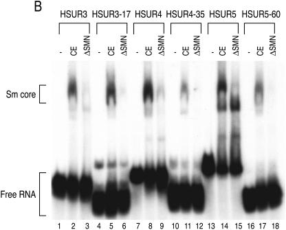
The survival of motor neurons (SMN) complex is essential for the biogenesis of spliceosomal small nuclear ribonucleoproteins (snRNPs) as it binds to and delivers Sm proteins for assembly of Sm cores on the abundant small nuclear RNAs (snRNAs). Using the conserved snRNAs encoded by the lymphotropic Herpesvirus saimiri (HVS), we determined the specific sequence and structural features of RNAs for binding to the SMN complex and for Sm core assembly. We show that the minimal SMN complex-binding domain in snRNAs, except U1, is comprised of an Sm site (AUUUUUG) and an adjacent 3' stem-loop. The adenosine and the first and third uridines of the Sm site are particularly critical for binding of the SMN complex, which directly contacts the backbone phosphates of these uridines. The specific sequence of the adjacent stem (7 to 12 base pairs)-loop (4 to 17 nucleotides) is not important for SMN complex binding, but it must be located within a short distance of the 3' end of the RNA for an Sm core to assemble. Importantly, these defining characteristics are discerned by the SMN complex and not by the Sm proteins, which can bind to and assemble on an Sm site sequence alone. These findings demonstrate that the SMN complex is the identifier, as well as assembler, of the abundant class of snRNAs in cells because it is able to recognize an snRNP code that they contain.


Figures

FIG. 1.
FIG. 1.
Mapping and direct binding of the minimal SMN complex-binding domains of the HSURs. (A) Native SMN complexes (SMN) were purified under high-salt conditions from stable cell lines expressing flag-Gemin2 (as described in Materials and Methods) and were analyzed by electrophoresis on 12.5% sodium dodecyl sulfate-polyacrylamide gels and silver staining. Immunoprecipitation using anti-flag antibody from the parental HeLa cell line was used as a control (Control). Core components of the SMN complex are labeled based upon molecular weight and Western blotting. Under these conditions, there is no detectable association of SMN complex with Sm proteins. (B) The SMN complex-binding domain of HSUR1. The 5′ (5′-P*)- and 3′ (3′-P*)-end-labeled HSUR1 was subjected to limited alkaline hydrolysis. The resulting hydrolyzed RNA ladders were incubated with purified SMN complex (SMN) or nonspecific proteins purified from HeLa cells (Control). The RNA fragments bound to the SMN complex were isolated and analyzed by electrophoresis on 7 M urea-8% acrylamide gels. The full-length RNA was digested with RNase T1 to provide a size marker. The solid red and blue arrows indicate the largest region that includes the SMN complex-binding domain, and open arrows indicate the smallest possible binding regions. For HSUR1, the open red arrow most likely indicates nonspecific degradation also seen in the input lane. Total represents 5% of input. (C) The SMN complex-binding domain of HSUR3. The same experiment as described in panel B was performed using 5′- and 3′-end-labeled HSUR3. The solid red and blue arrows indicate the largest region that includes the SMN complex-binding domain, and open arrows indicate the smallest possible binding regions. (D) The 5′ end deletion mutants of HSUR1 were transcribed in the presence of [32P]UTP and incubated with flag-purified SMN complex or nonspecific HeLa control proteins (control) for 1 h at 4°C. Bound RNAs were isolated and analyzed by electrophoresis on 7 M urea-8% polyacrylamide gels and autoradiography. Total represents 10% of input. (E) The 5′ end deletion mutants of HSUR3 were transcribed in the presence of [32P]UTP and subjected to SMN complex binding as described in panel D. (F) The 5′-end deletion mutants of HSUR4 were transcribed in the presence of [32P]UTP and subjected to SMN complex binding as described in panel D. Control not shown. (G) The 5′-end deletion mutants of HSUR5 were transcribed in the presence of [32P]UTP and subjected to SMN complex binding as described in panel D. Control not shown.
FIG. 1.
FIG. 1.
Mapping and direct binding of the minimal SMN complex-binding domains of the HSURs. (A) Native SMN complexes (SMN) were purified under high-salt conditions from stable cell lines expressing flag-Gemin2 (as described in Materials and Methods) and were analyzed by electrophoresis on 12.5% sodium dodecyl sulfate-polyacrylamide gels and silver staining. Immunoprecipitation using anti-flag antibody from the parental HeLa cell line was used as a control (Control). Core components of the SMN complex are labeled based upon molecular weight and Western blotting. Under these conditions, there is no detectable association of SMN complex with Sm proteins. (B) The SMN complex-binding domain of HSUR1. The 5′ (5′-P*)- and 3′ (3′-P*)-end-labeled HSUR1 was subjected to limited alkaline hydrolysis. The resulting hydrolyzed RNA ladders were incubated with purified SMN complex (SMN) or nonspecific proteins purified from HeLa cells (Control). The RNA fragments bound to the SMN complex were isolated and analyzed by electrophoresis on 7 M urea-8% acrylamide gels. The full-length RNA was digested with RNase T1 to provide a size marker. The solid red and blue arrows indicate the largest region that includes the SMN complex-binding domain, and open arrows indicate the smallest possible binding regions. For HSUR1, the open red arrow most likely indicates nonspecific degradation also seen in the input lane. Total represents 5% of input. (C) The SMN complex-binding domain of HSUR3. The same experiment as described in panel B was performed using 5′- and 3′-end-labeled HSUR3. The solid red and blue arrows indicate the largest region that includes the SMN complex-binding domain, and open arrows indicate the smallest possible binding regions. (D) The 5′ end deletion mutants of HSUR1 were transcribed in the presence of [32P]UTP and incubated with flag-purified SMN complex or nonspecific HeLa control proteins (control) for 1 h at 4°C. Bound RNAs were isolated and analyzed by electrophoresis on 7 M urea-8% polyacrylamide gels and autoradiography. Total represents 10% of input. (E) The 5′ end deletion mutants of HSUR3 were transcribed in the presence of [32P]UTP and subjected to SMN complex binding as described in panel D. (F) The 5′-end deletion mutants of HSUR4 were transcribed in the presence of [32P]UTP and subjected to SMN complex binding as described in panel D. Control not shown. (G) The 5′-end deletion mutants of HSUR5 were transcribed in the presence of [32P]UTP and subjected to SMN complex binding as described in panel D. Control not shown.
FIG.2.
FIG.2.
The minimal SMN complex-binding domains are sufficient for SMN-dependent Sm core assembly. (A) The secondary structures of HSUR1, HSUR3, HSUR4, and HSUR5 and their SMN complex-binding domains (highlighted in pink). Solid blue and red arrows designate the maximum 5′ and 3′ end boundaries of the SMN complex-binding domains, and open blue arrows indicate smaller domains that mediate weak binding to the SMN complex. The open red arrow indicates nonspecific degradation. (B) [32P]UTP-labeled HSUR3, HSUR3-17, HSUR4, HSUR4-35, HSUR5, and HSUR5-60 were incubated with buffer (−), HeLa mock-depleted cytoplasmic extracts (CE), or SMN complex-depleted HeLa extracts (ΔSMN) for 1 h at 30°C. The assembly reaction products were analyzed by electrophoresis on 6% native polyacrylamide gels and autoradiography. Sm cores and free RNAs are each indicated by brackets.
FIG.2.
FIG.2.
The minimal SMN complex-binding domains are sufficient for SMN-dependent Sm core assembly. (A) The secondary structures of HSUR1, HSUR3, HSUR4, and HSUR5 and their SMN complex-binding domains (highlighted in pink). Solid blue and red arrows designate the maximum 5′ and 3′ end boundaries of the SMN complex-binding domains, and open blue arrows indicate smaller domains that mediate weak binding to the SMN complex. The open red arrow indicates nonspecific degradation. (B) [32P]UTP-labeled HSUR3, HSUR3-17, HSUR4, HSUR4-35, HSUR5, and HSUR5-60 were incubated with buffer (−), HeLa mock-depleted cytoplasmic extracts (CE), or SMN complex-depleted HeLa extracts (ΔSMN) for 1 h at 30°C. The assembly reaction products were analyzed by electrophoresis on 6% native polyacrylamide gels and autoradiography. Sm cores and free RNAs are each indicated by brackets.
FIG. 3.
FIG. 3.
A terminal stem-loop is necessary for SMN complex binding in vitro and Sm core assembly in vivo. (A) Illustration of HSUR5-60 RNAs used in this experiment. Site-directed mutagenesis was used to destabilize the terminal stem of HSUR5-60 (des-stem) and either 15 or 70 nt were added to the 3′ end of HSUR5-60 (+15 nt and +70 nt). (B) HSUR5-60 wild type (WT) or des-stem RNAs were labeled at the 5′ end with [γ-32P]ATP and incubated with serial dilutions of RNase T1 for 15 min at 25°C. The digested RNAs were purified and analyzed by electrophoresis on 7 M urea-12% polyacrylamide gels and autoradiography. (C) HSUR5-60 (WT), des-stem, +15-nt, and +70-nt RNAs or an HSUR5-60 RNA with a complete deletion of the 3′ stem-loop (no-stem) were [32P]UTP labeled, mixed with U6 as a negative control, and incubated with purified SMN complex for 1 h at 4°C. Bound RNAs were isolated and analyzed by electrophoresis on 7 M urea-8% polyacrylamide gels and autoradiography. Total represents 10% of input. (D) Experimental strategy used for Xenopus oocyte microinjection. (E) HSUR5-60 (WT), des-stem (the lower band), or +15 nt and +70 nt RNAs were [32P]UTP labeled, mixed with labeled U6, and injected into the cytoplasm of oocytes as shown in panel D. After incubation for 1.5 h, the oocytes were homogenized, and immunoprecipitations were carried out with either anti-Sm (Y12) or control nonimmune (SP2/0) antibodies. RNAs were purified and analyzed by electrophoresis on 7 M urea-8% polyacrylamide gels. Total represents 10% of the fractions used for each immunoprecipitation. The levels of assembly were quantitated using imaging software and normalized to the wild type.
FIG. 3.
FIG. 3.
A terminal stem-loop is necessary for SMN complex binding in vitro and Sm core assembly in vivo. (A) Illustration of HSUR5-60 RNAs used in this experiment. Site-directed mutagenesis was used to destabilize the terminal stem of HSUR5-60 (des-stem) and either 15 or 70 nt were added to the 3′ end of HSUR5-60 (+15 nt and +70 nt). (B) HSUR5-60 wild type (WT) or des-stem RNAs were labeled at the 5′ end with [γ-32P]ATP and incubated with serial dilutions of RNase T1 for 15 min at 25°C. The digested RNAs were purified and analyzed by electrophoresis on 7 M urea-12% polyacrylamide gels and autoradiography. (C) HSUR5-60 (WT), des-stem, +15-nt, and +70-nt RNAs or an HSUR5-60 RNA with a complete deletion of the 3′ stem-loop (no-stem) were [32P]UTP labeled, mixed with U6 as a negative control, and incubated with purified SMN complex for 1 h at 4°C. Bound RNAs were isolated and analyzed by electrophoresis on 7 M urea-8% polyacrylamide gels and autoradiography. Total represents 10% of input. (D) Experimental strategy used for Xenopus oocyte microinjection. (E) HSUR5-60 (WT), des-stem (the lower band), or +15 nt and +70 nt RNAs were [32P]UTP labeled, mixed with labeled U6, and injected into the cytoplasm of oocytes as shown in panel D. After incubation for 1.5 h, the oocytes were homogenized, and immunoprecipitations were carried out with either anti-Sm (Y12) or control nonimmune (SP2/0) antibodies. RNAs were purified and analyzed by electrophoresis on 7 M urea-8% polyacrylamide gels. Total represents 10% of the fractions used for each immunoprecipitation. The levels of assembly were quantitated using imaging software and normalized to the wild type.
FIG. 4.
FIG. 4.
A terminal stem-loop is required for SMN-dependent Sm core assembly. (A) A total of 10,000 cpm of HSUR5-60 (WT), des-stem, or +15-nt and +70-nt RNAs (Fig. 3) were incubated with HeLa snRNP TPs for 1 h at 30°C and then subjected to immunoprecipitation with anti-Sm (Y12) antibody. RNAs with assembled Sm cores were isolated and analyzed by electrophoresis on 7 M urea-8% polyacrylamide gels and autoradiography. The levels of assembly were quantitated using imaging software and normalized to the wild type, and the input values were normalized prior to assembly. The composition of TPs is shown in panel E. (B) The RNAs used in panel A were incubated with low-salt-purified SMN complex for 1 h at 30°C and then subjected to immunoprecipitation with anti-Sm (Y12) antibody. RNAs with assembled Sm cores were isolated and analyzed by electrophoresis on 7 M urea-8% polyacrylamide gels and autoradiography. The levels of assembly were quantitated using imaging software and normalized to the wild type, and the input values were normalized prior to assembly. The composition of SMN complex is shown in panel D. (C) HSUR5-60 no-stem (no 3′ stem-loop) RNA was incubated with low-salt-purified SMN complex (SMN) or TPs for 1 h at 30°C and then subjected to immunoprecipitation with anti-Sm (Y12) antibody. RNAs with assembled Sm cores were isolated and analyzed by electrophoresis on 7 M urea-8% polyacrylamide gels and autoradiography. Input represents 10% of total. The experiments shown in panels B and C were performed in parallel. (D) Native SMN complexes (SMN) or nonspecific proteins (Control) were purified from flag-Gemin2 cells or the parental HeLa cells, respectively, under low-salt conditions as described in Materials and Methods. Flag-purified proteins were eluted with 3× Flag peptide, resolved by electrophoresis on 4 to 12% gradient polyacrylamide gels, and analyzed by silver staining. Under these conditions, all seven Sm proteins copurify with the SMN complex. (E) Native snRNP TPs were purified from HeLa cells as described in Materials and Methods and were analyzed by electrophoresis on 4 to 12% gradient polyacrylamide gels and by silver staining.
FIG. 5.
FIG. 5.
The sequence of the 3′ stem-loop is not critical for SMN complex binding in vitro or Sm core assembly in vivo. (A) Illustration of the constructs used in this experiment. (B) The wild-type (WT), flip, swap, and loop constructs of HSUR4-35 and HSUR5-60, as illustrated in panel A, were labeled with [32P]UTP, mixed with U6 as a negative control, and incubated with purified SMN complex for 1 h at 4°C. Bound RNAs were isolated and analyzed by electrophoresis on 7 M urea-8% polyacrylamide gels and autoradiography. The wild type has a different electrophoretic mobility than the flip, swap, and loop constructs due to digestion with a different restriction enzyme prior to runoff transcription. Total represents 10% of input. (C) The wild-type, flip, swap, and loop constructs of HSUR4-35 were labeled with [32P]UTP, mixed with labeled U6, and injected into the cytoplasm of Xenopus oocytes. After a 1.5-h incubation, the oocytes were homogenized, and immunoprecipitations were carried out with either anti-Sm (Y12) or control nonimmune (SP2/0) antibodies. RNAs were purified and analyzed by electrophoresis on 7 M urea-8% polyacrylamide gels. Total represents 10% of the fractions used for each immunoprecipitation. (D) The wild-type, flip, swap, and loop constructs of HSUR5-60 were subjected to the same experiment as described in panel C.
FIG. 6.
FIG. 6.
The first and third uridines of the Sm site are essential for SMN complex binding (and Sm core assembly in vitro). (A) Wild-type HSUR5-60 (AUUUUUG) or HSUR5-60 in which each position of the Sm site was changed one at a time to either a guanosine or a cytosine (Sm site substitutions are indicated in red) was [32P]UTP labeled, mixed with U6 as a negative control, and incubated with purified SMN complex (SMN complex) or control HeLa proteins (Control) for 1 h at 4°C. Bound RNAs were isolated and analyzed by electrophoresis on 7 M urea-8% polyacrylamide gels and by autoradiography. Total represents 10% of input. The levels of binding were quantitated using Photoshop and normalized to the wild type. (B) A subset of the RNAs used in panel A were incubated with low-salt-purified SMN complex (Fig. 4D) for 1 h at 30°C and then subjected to immunoprecipitation with anti-Sm (Y12) antibody. RNAs with assembled Sm cores were isolated and analyzed by electrophoresis on 7 M urea-8% polyacrylamide gels and autoradiography. (C) The RNAs used in panel B were incubated with TPs (Fig. 4E) for 1 h at 30°C and then subjected to immunoprecipitation with anti-Sm (Y12) antibody. RNAs with assembled Sm cores were isolated and analyzed by electrophoresis on 7 M urea-8% polyacrylamide gels and autoradiography.
FIG. 7.
FIG. 7.
The SMN complex contacts the phosphate backbone of the first and third uridines of the Sm site.(A) Phosphorothioate interference of HSUR5-60. HSUR5-60 transcribed in the presence of A, U, G, or C α-thiotriphosphate or no analog (−) was 5′ end labeled with [γ-32P]ATP and incubated with purified SMN complex for 1 h at 4°C. Bound RNAs were isolated and cleaved with iodine, and the resulting fragments were resolved on a 7 M urea-12% polyacrylamide sequencing gel. Unbound (Total) RNA was also cleaved with iodine to correct for phosphorothioate incorporation and cleavage efficiency. The Sm site is indicated by brackets. Solid red circles to the right of U76 and U78 indicate positions where the phosphorothioate substitution interferes with SMN complex binding. (B) Phosphorothioate interference of HSUR4-35. The same experiment as described in panel A was performed using 5′-end-labeled HSUR4-35. The cleaved RNAs were resolved on a 7 M urea-10% polyacrylamide sequencing gel. Solid red circles to the right of U71, U73, U89, and C65 indicate positions where the phosphorothioate substitution interferes with SMN complex binding. (C) Kappa values for phosphorothioate interference at HSUR5-60 uridines. Sm site uridines are numbered in red. An increased κ value is interpreted as an interference. (D) Kappa values for phosphorothioate interference at HSUR4-35 uridines. Sm site uridines are numbered in red. An increased κ value is interpreted as an interference.
FIG. 8.
FIG. 8.
Sm proteins do not cause phosphorothioate interference at Sm site uridines. (A) HSUR5-60 transcribed in the presence A, U, G, or C α-thiotriphosphate or no analog (−) was 5′ end labeled with [γ-32P]ATP and incubated with purified TPs (Fig. 4E) for 1 h at 30°C. Bound RNAs were isolated by immunoprecipitation with anti-Sm (Y12) antibody and cleaved with iodine, and the resulting fragments were resolved on a 7 M urea-12% polyacrylamide sequencing gel. Unbound (Total) RNA was also cleaved with iodine to correct for phosphorothioate incorporation and cleavage efficiency. The Sm site is indicated by brackets. Solid blue circles to the right of the specified bands indicate positions where the phosphorothioate substitution interferes with TP binding and assembly. The adenosines (A) can be seen with increased exposure. (B) Kappa values for phosphorothioate interference at HSUR5-60 uridines. Sm site uridines are numbered in red. An increased κ value is interpreted as an interference. (C) Secondary structure of the region of HSUR5-60 used for phosphorothioate interference. Blue arrows indicate positions where phosphorothioate substitution interferes with TP binding and assembly. (D) HeLa cell extracts (Total), high-salt-purified SMN complex (SMN complex), and snRNP total proteins (TPs) were run on 4 to 12% gradient polyacrylamide gels and Western blotted for components of the SMN complex.
FIG. 9.
FIG. 9.
Model for the critical RNA sequence features that confer binding to the SMN complex and assembly of an Sm core. The details of these features are described in the text.

Similar articles

Cited by

References

    1. Baccon, J., L. Pellizzoni, J. Rappsilber, M. Mann, and G. Dreyfuss. 2002. Identification and characterization of Gemin7, a novel component of the survival of motor neuron complex. J. Biol. Chem. 277:31957-31962. - PubMed
    1. Brahms, H., Lo. Meheus, V. de Brabandere, U. Fischer, and R. Luhrmann. 2001. Symmetrical dimethylation of arginine residues in spliceosomal Sm protein B/B′ and the Sm-like protein LSm4, and their interaction with the SMN protein. RNA 7:1531-1542. - PMC - PubMed
    1. Branlant, C., A. Krol, J. P. Ebel, E. Lazar, B. Haendler, and M. Jacob. 1982. U2 RNA shares a structural domain with U1, U4, and U5 RNAs. EMBO J. 1:1259-1265. - PMC - PubMed
    1. Buhler, D., V. Raker, R. Luhrmann, and U. Fischer. 1999. Essential role for the tudor domain of SMN in spliceosomal U snRNP assembly: implications for spinal muscular atrophy. Hum. Mol. Genet. 8:2351-2357. - PubMed
    1. Charroux, B., L. Pellizzoni, R. A. Perkinson, A. Shevchenko, M. Mann, and G. Dreyfuss. 1999. Gemin3: A novel DEAD box protein that interacts with SMN, the spinal muscular atrophy gene product, and is a component of gems. J. Cell Biol. 147:1181-1194. - PMC - PubMed

Publication types

MeSH terms

LinkOut - more resources