. 2005 Dec;25(24):11113-21.
doi: 10.1128/MCB.25.24.11113-11121.2005.

Essential role for sphingosine kinases in neural and vascular development

Affiliations

Essential role for sphingosine kinases in neural and vascular development

Kiyomi Mizugishi et al. Mol Cell Biol. 2005 Dec.

Abstract

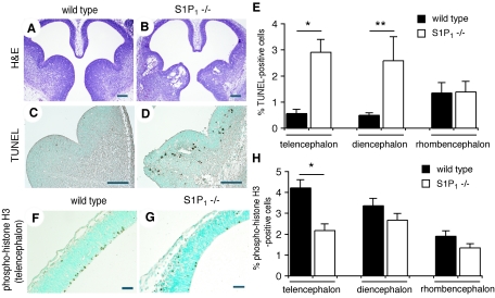
Sphingosine-1-phosphate (S1P), an important sphingolipid metabolite, regulates diverse cellular processes, including cell survival, growth, and differentiation. Here we show that S1P signaling is critical for neural and vascular development. Sphingosine kinase-null mice exhibited a deficiency of S1P which severely disturbed neurogenesis, including neural tube closure, and angiogenesis and caused embryonic lethality. A dramatic increase in apoptosis and a decrease in mitosis were seen in the developing nervous system. S1P(1) receptor-null mice also showed severe defects in neurogenesis, indicating that the mechanism by which S1P promotes neurogenesis is, in part, signaling from the S1P(1) receptor. Thus, S1P joins a growing list of signaling molecules, such as vascular endothelial growth factor, which regulate the functionally intertwined pathways of angiogenesis and neurogenesis. Our findings also suggest that exploitation of this potent neuronal survival pathway could lead to the development of novel therapeutic approaches for neurological diseases.


Figures

FIG. 1.
FIG. 1.
Targeted disruption of the SphK2 gene. (A) Schematic representation of the SphK2 targeting strategy. The structure of the SphK2 targeting vector is shown at the top, the mouse SphK2 locus in the middle, and the predicted structure of the homologous recombined locus, with the location of the Southern probe, at the bottom. P1 and P2 were used for RT-PCR. B, BamHI; Bg, BglII; N, NotI; S, SpeI; X, XhoI; pBS; pBluescript vector. (B) Southern blot analysis of SpeI-digested genomic tail DNA from mice generated by SphK2 heterozygous mating. The wild-type SphK2 locus yielded a 7.0-kb SpeI band. The disrupted SphK2 locus yielded a 10.9-kb SpeI band. (C) RT-PCR analysis of total RNA from adult tissues with primers P1 and P2. SphK2+/+ RNA yielded the predicted 195-bp amplification product. No amplification product was detected from SphK2−/− RNA. GAPDH, glyceraldehyde-3-phosphate dehydrogenase.
FIG. 2.
FIG. 2.
Measurement of SphK activity and S1P levels in SphK1 SphK2 combined mutants. (A and B) SphK enzymatic activity was determined in wild-type and SphK1−/− SphK2+/+, SphK1+/+ SphK2−/−, and SphK1−/− SphK2−/− mutant embryos at E11.5. The assay was performed in the presence of Triton X-100 (A) or in BSA complexes without Triton X-100 (B). The data represent means ± SEs (n = 3; *, P < 0.01; **, P < 0.05 [paired t test compared with the wild type]). (C) S1P levels in wild-type and SphK1−/− SphK2+/+, SphK1+/+ SphK2−/−, and SphK1−/− SphK2−/− mutant embryos at E11.5. The data represent means ± SEs (n = 3; **, P < 0.05 [paired t test compared with the wild type]) (D) Sphingosine levels in wild-type and SphK1−/− SphK2−/− mutant embryos at E11.5. The data represent means ± SEs (n = 3).
FIG. 3.
FIG. 3.
Vascular defects in SphK1−/− SphK2−/− mutants. (A to D) Photomicrographs of E11.5 embryos. SphK1−/− SphK2−/− mutant embryos show hemorrhages in the brain (B to D), mandible (B), and spinal cord (C), sometimes along with enlargement of the cardiac profile (D). (E to H) H&E staining of sagittal sections from SphK1−/− SphK2−/− mutant embryos at E11.5. (E and F) Hemorrhage in the cranial mesenchymal region (arrows). (G) Intraventricular hemorrhage (arrow). (H) Pericardial hemorrhage (arrows). FV, fourth ventricle; Ht, heart; LV, lateral ventricle; Mes, mesenchyme; NE, neuroepithelium; TE, telencephalon; RE, rhombencephalon. (I to L) Vascular smooth muscle defects in SphK1−/− SphK2−/− mutants at E11.5. (I and K) H&E staining of sagittal sections of the dorsal aorta. The arrows in panel I and K indicate the wall of the aorta. (J and L) Transverse sections of the dorsal aorta stained with anti-smooth muscle α-actin (SMαA). (M to P) Electron microscopic analysis of blood vessels from the capillaries in the cranial mesenchymal region from E11.5 wild-type (M and N) and SphK1−/− SphK2−/− mutant (O and P) embryos. The arrows in panel O indicate broken endothelial cells. BC, blood cell; EC, endothelial cell; EJ, endothelial cell-cell junction; PC, pericyte; Vac, vacuole. Magnifications: M, ×1,600; N, ×50,000; O and P, ×2,000. (Q to T) Whole-mount immunostaining with anti-PECAM-1 antibody of blood vessels in the brains of E10.5 wild-type (Q and R) and SphK1−/− SphK2−/− mutant (S and T) embryos. Note the dilated blood vessels (arrows in panel S) and aberrant network (arrowheads in panel T). Scale bars represent 200 μm (E, F, G, H, I, and K) and 50 μm (J and L).
FIG. 4.
FIG. 4.
NTDs in SphK1−/− SphK2−/− mutants. (A to D) Exencephaly in SphK1−/− SphK2−/− mutant embryos. (A and B) Wild-type control embryos at E10.5. (C and D) SphK1−/− SphK2−/− mutant embryos at E10.5. (A and C) Lateral view. (B and D) Dorsal view. Ex, exencephaly. (E to H) Expression of SphK1, SphK2, and S1P1 in E10.5 embryos. (E) Whole-mount in situ hybridization for SphK1 in a wild-type embryo. (F, G, and H) Whole-mount X-Gal staining of SphK2/ and S1P1/ mutant and wild-type embryos, respectively. The lacZ reporter gene was inserted into the SphK2 and S1P1 loci. TE, telencephalon.
FIG. 5.
FIG. 5.
Increased cell death and decreased cell proliferation in SphK1−/− SphK2−/− mutants. (A and B) H&E staining of transverse sections from wild-type (A) and SphK1−/− SphK2−/− mutant (B) embryos at E9.5. An arrow in panel B indicates an NTD. (C and D) TUNEL assay of wild-type (C) and SphK1−/− SphK2−/− mutant (D) embryos at E9.5. The boxed areas in panels A and B are shown, respectively. NE, neuroepithelium; Mes, mesenchyme. (E to H) H&E staining of sections from wild-type (E and G) and SphK1−/− SphK2−/− mutant (F and H) embryos at E11.5. (E and F) Sagittal sections. (G and H) Transverse sections. DE, diencephalon; FV, fourth ventricle; ME, mesencephalon; RE, rhombencephalon; TE, telencephalon; TV, third ventricle. (I and J) TUNEL assay of wild-type (I) and SphK1−/− SphK2−/− mutant (J) embryos at E11.5. The telencephalon is shown. (K) Percentage of TUNEL-positive cells (n = 5 matched pairs; *, P < 0.01 [paired t test]). (L and M) Immunostaining with anti-phospho-histone H3 of wild-type (L) and SphK1−/− SphK2−/− mutant (M) embryos at E11.5. The telencephalon is shown. (N) Percentage of phospho-histone H3-positive cells (n = 4 matched pairs; *, P < 0.01 [paired t test]). Scale bars represent 500 μm (A, B, E, F, G, and H) and 50 μm (C, D, I, J, L, and M).
FIG. 6.
FIG. 6.
Requirement of S1P1 receptor for neural development. (A and B) H&E staining of transverse sections from wild-type (A) and S1P1−/− mutant (B) embryos at E12.5. (C and D) TUNEL assay of wild-type (C) and S1P1−/− mutant (D) embryos at E12.5. (E) Percentage of TUNEL-positive cells (n = 6 matched pairs; *, P < 0.01; **, P < 0.05 [paired t test]). (F and G) Immunostaining with anti-phospho-histone H3 of wild-type (F) and S1P1−/− mutant (G) embryos at E12.5. (H) Percentage of phospho-histone H3-positive cells (n = 6 matched pairs; *, P < 0.01 [paired t test]). Scale bars represent 200 μm (A, B, C, and D) and 50 μm (F and G).

References

    1. Allende, M. L., T. Yamashita, and R. L. Proia. 2003. G-protein-coupled receptor S1P1 acts within endothelial cells to regulate vascular maturation. Blood 102:3665-3667. - PubMed
    1. Allende, M. L., J. L. Dreier, S. Mandala, and R. L. Proia. 2004. Expression of the sphingosine-1-phosphate receptor, S1P1, on T-cells controls thymic emigration. J. Biol. Chem. 279:15396-15401. - PubMed
    1. Allende, M. L., T. Sasaki, H. Kawai, A. Olivera, Y. Mi, G. van Echten-Deckert, R. Hajdu, M. Rosenbach, C. A. Keohane, S. Mandala, S. Spiegel, and R. L. Proia. 2004. Mice deficient in sphingosine kinase 1 are rendered lymphopenic by FTY720. J. Biol. Chem. 279:52487-52492. - PubMed
    1. Bamforth, S. D., J. Bragança, J. Eloranta, J. N. Murdoch, F. I. Marques, K. R. Kranc, H. Farza, D. J. Henderson, H. C. Hurst, and S. Bhattacharya. 2001. Cardiac malformations, adrenal agenesis, neural crest defects and exencephaly in mice lacking Cited2, a new Tfap2 co-activator. Nat. Genet. 29:469-474. - PubMed
    1. Cinamon, G., M. Matloubian, M. J. Lesneski, Y. Xu, C. Low, T. Lu, R. L. Proia, and J. G. Cyster. 2004. Sphingosine 1-phosphate receptor 1 promotes B cell localization in the splenic marginal zone. Nat. Immunol. 5:713-720. - PubMed

Publication types

MeSH terms