. 2005 Dec 6;102(49):17565-9.
doi: 10.1073/pnas.0506010102. Epub 2005 Nov 28.

Breaking the diffraction barrier in fluorescence microscopy at low light intensities by using reversibly photoswitchable proteins

Affiliations

Breaking the diffraction barrier in fluorescence microscopy at low light intensities by using reversibly photoswitchable proteins

Michael Hofmann et al. Proc Natl Acad Sci U S A. .

Abstract

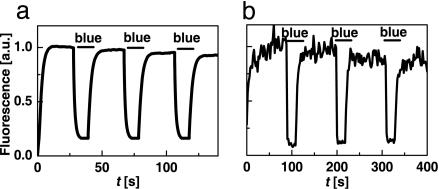
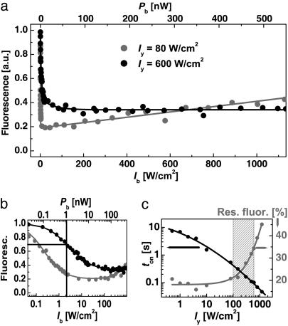
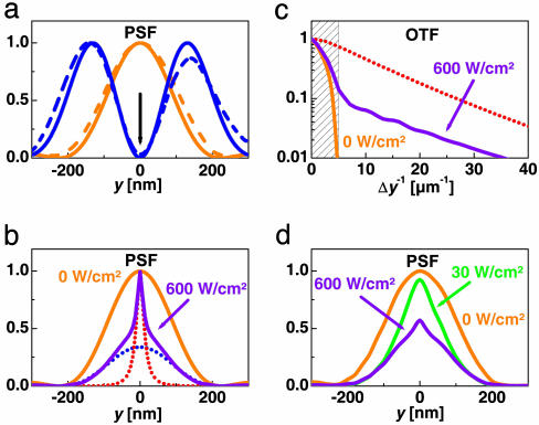
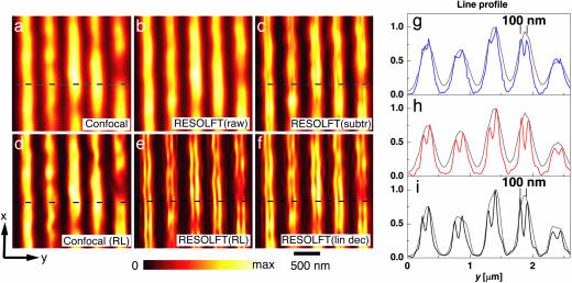
Fluorescence microscopy is indispensable in many areas of science, but until recently, diffraction has limited the resolution of its lens-based variant. The diffraction barrier has been broken by a saturated depletion of the marker's fluorescent state by stimulated emission, but this approach requires picosecond laser pulses of GW/cm2 intensity. Here, we demonstrate the surpassing of the diffraction barrier in fluorescence microscopy with illumination intensities that are eight orders of magnitude smaller. The subdiffraction resolution results from reversible photoswitching of a marker protein between a fluorescence-activated and a nonactivated state, whereby one of the transitions is accomplished by means of a spatial intensity distribution featuring a zero. After characterizing the switching kinetics of the used marker protein asFP595, we demonstrate the current capability of this RESOLFT (reversible saturable optical fluorescence transitions) type of concept to resolve 50-100 nm in the focal plane. The observed resolution is limited only by the photokinetics of the protein and the perfection of the zero. Our results underscore the potential to finally achieve molecular resolution in fluorescence microscopy by technical optimization.


Figures

Fig. 1.
Fig. 1.
Wide-field photoswitching of asFP595 fluorescence from an E. coli colony (a) and on a 0.3-μm-diameter focal spot of an ultrathin asFP595 layer (b). The fluorescence is elicited by 10 mW (a) and 3.3 nW (b) of continuous wave irradiation with yellow light. Inhibition results from addition of 0.3 mW (a) and 2.2 nW (b) of blue light.
Fig. 2.
Fig. 2.
Characterization of asFP595 photoswitching. (a) Inhibition of fluorescence as a function of the blue intensity Ib for the yellow intensities Iy = 80 W/cm2 (black circles) and 600 W/cm2 (gray circles). (b) Same data on a semilogarithmic plot to disclose Isat.(c) Effect of Iy on the time ton needed for settling the fluorescence level (black circles) as well as on the minimal residual fluorescence after inhibition (gray circles).
Fig. 3.
Fig. 3.
Subdiffraction point spread and transfer function by asFP595 photoswitching. (a) Measured and calculated In-PSF (blue) with the PSF for fluorescence generation (yellow). Solid lines are calculated, and dashed lines are experimental. (b) E-PSF calculated for Iy = 600 W/cm2 and different intensities Ib = 600 W/cm2 (violet) and Ib = 0 (yellow). In the former case, the E-PSF consists of a diffraction-limited PSFdiff (blue dots) plus a subdiffraction PSFRESOLFT counterpart (red dots) (ε = 0.3, Eq. 2). PSFRESOLFT is attained as well when disregarding the action cross-talk of blue light within the calculations. (c) The associated modulus of the effective OTF (violet) and OTFRESOLFT (red) quantifies the gain in spatial frequency bandwidth over the diffraction limit (shaded) or reference OTF for Ib = 0 (yellow). (d) E-PSFs calculated in the same way as in b but employing the experimental In-PSFs.
Fig. 4.
Fig. 4.
Subdiffraction fluorescence microscopy with asFP595. Nanofabricated grooves stained with asFP595 and imaged without (a) and with (b) the blue In-PSF. c shows the weighted subtraction of a from b. d exhibits the restored data of a; e is its counterpart based on b. f shows the linearly deconvolved image of b. g compares line profiles from the images a and c, extracted at the dashed horizontal line, in gray and blue, respectively. h shows the analogous comparison for a and f; i compares the profiles for d and e. The 35% depth of the 100-nm distant peaks of the linearly deconvolved data in h enable an extrapolation of resolution of distances <100 nm. The sharp peaks are a hallmark of the saturation process and of RESOLFT microscopy in general.

References

    1. Fraser, S. E. (2003) Nat. Biotechnol. 21, 1272–1273. - PubMed
    1. Tsien, R. Y. (2003) Nat. Cell Biol., SS16–SS21. - PubMed
    1. Abbe, E. (1873) Arch. Mikr. Anat. 9, 413–420.
    1. Hell, S. W., Jakobs, S. & Kastrup, L. (2003) Appl. Phys. A 77, 859–860.
    1. Hell, S. W. (2003) Nat. Biotechnol. 21, 1347–1355. - PubMed

Publication types

Substances