. 2006 Oct;55(10):1238-46.
doi: 10.1007/s00262-005-0104-8. Epub 2005 Nov 29.

Induction of anti-tumor immunity by vaccination with dendritic cells pulsed with anti-CD44 IgG opsonized tumor cells

Affiliations

Induction of anti-tumor immunity by vaccination with dendritic cells pulsed with anti-CD44 IgG opsonized tumor cells

Shari Pilon-Thomas et al. Cancer Immunol Immunother. 2006 Oct.

Abstract

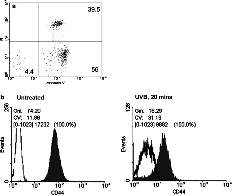
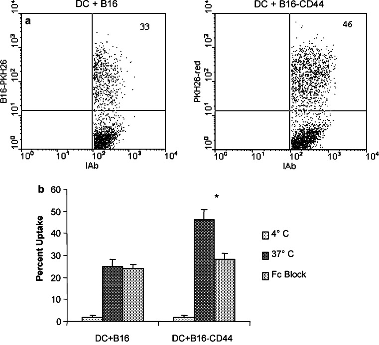
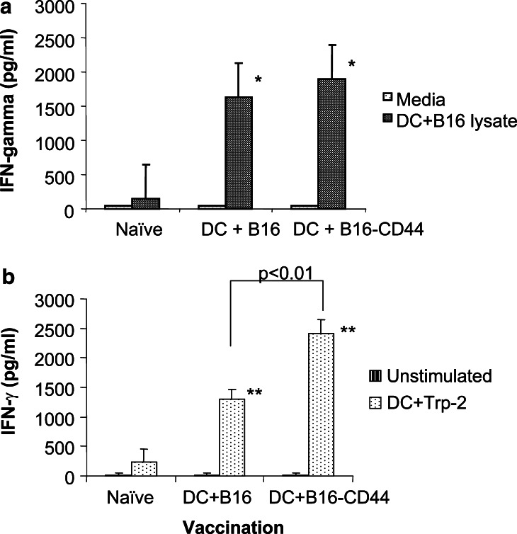
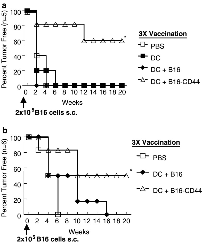
Due to the pivotal role that dendritic cells (DC) play in eliciting and maintaining functional anti-tumor T cell responses, these APC have been exploited against tumors. DC express several receptors for the Fc portion of IgG (Fcgamma receptors) that mediate the internalization of antigen-IgG complexes and promote efficient MHC class I and II restricted antigen presentation. In this study, the efficacy of vaccination with DC pulsed with apoptotic B16 melanoma cells opsonized with an anti-CD44 IgG (B16-CD44) was explored. Immature bone marrow derived DC grown in vitro with IL-4 and GM-CSF were pulsed with B16-CD44. After 48 h of pulsing, maturation of DC was demonstrated by production of IL-12 and upregulation of CD80 and CD40 expression. To test the efficacy of vaccination with DC+B16-CD44, mice were vaccinated subcutaneously Lymphocytes from mice vaccinated with DC+B16-CD44 produced IFN-gamma in response to B16 melanoma lysates as well as an MHC class I restricted B16 melanoma-associated peptide, indicating B16 specific CD8 T cell activation. Upon challenge with viable B16 cells, all mice vaccinated with DC alone developed tumor compared to 40% of mice vaccinated with DC+B16-CD44; 60% of the latter mice remained tumor free for at least 8 months. In addition, established lung tumors and distant metastases were significantly reduced in mice treated with DC+B16-CD44. Lastly, delayed growth of established subcutaneous tumors was induced by combination therapy with anti-CD44 antibodies followed by DC injection. This study demonstrates the efficacy of targeting tumor antigens to DC via Fcgamma receptors.


Figures

Fig. 1
Fig. 1
Phenotype and viability of B16 melanoma after UVB exposure. a B16 cells (1×10⁶/ml PBS) were exposed to UVB light for 20 min. Cells were stained with Annexin V and PI for determination of apoptotic and necrotic cell population by FACS, b CD44 expression on B16 cells was measured by FACS prior to and after UVB exposure. Filled histograms indicate CD44 positive cells while the negative control is shown as empty histograms
Fig. 2
Fig. 2
Binding of B16 cells by DC via Fcγ Receptors. a B16 cells were labeled with PKH26 red dye and treated for 20 min with UVB irradiation. Cells were coated with normal rat IgG or anti-CD44 antibodies prior to a 24 h incubation with day 5 DC. DC were stained for MHC class II expression. Two-color analysis revealed cells that were positive for both PKH26 red dye and MHC class II, indicative of DC that have taken up tumor cells, b Prior to co-culture with UVB treated, PKH26 red labeled B16 cells, DC were pretreated with anti-CD16/CD32 antibodies (Fc block) to block Fcγ receptors, * indicates p<0.05
Fig. 3
Fig. 3
Maturation of DC after uptake of B16 cells. DC were co-cultured for 24 h with UVB treated B16 cells stained with normal rat IgG (DC + B16) or anti-CD44 antibodies (DC + B16-CD44). a Expression of CD86, CD80, and CD40 was measured on the surface of MHC class II positive cells by FACS. Dotted histogram = negative control, gray histogram = DC alone, black histogram = DC + B16, filled histogram = DC + B16-CD44, b Supernatants were collected and IL-12p70 was measured by standard ELISA
Fig. 4
Fig. 4
IFN-gamma production in response to B16 lysate and peptide. Mice were vaccinated three times at 2 week intervals with DC + B16 or DC + B16-CD44. One week after the final vaccination, lymph node cells were collected and co-cultured for 48 h with a DC + B16 lysate or b Trp-2 peptide. IFN-g production was measured by standard ELISA. Data are shown as mean ± SEM of two independent experiments. * indicates p<0.05 and ** indicates p<0.01 compared to naïve lymph node cells
Fig. 5
Fig. 5
Protection against B16 tumor. a Mice (n=5) were vaccinated s.c. three times at 2 week intervals with PBS alone, DC alone, or DC pulsed for 24 h with UVB treated B16 cells coated with normal rat IgG (DC + B16) or anti-CD44 antibodies (DC + B16-CD44). Two weeks after the final vaccination, mice were challenged with 2×10⁵ B16 cells in the opposite flank. The results of three experiments were very similar and are therefore combined in this figure, b Mice (n=6) were vaccinated s.c. three times at 2 week intervals with PBS alone, or DC pulsed for 24 h with UVB treated B16 cells coated with normal rat IgG (DC + B16) or anti-CD44 antibodies (DC + B16-CD44). Prior to injection, CD11c+ DC were purified by MACS separation. Two weeks after the final vaccination, mice were challenged with 2×10⁵ B16 cells in the opposite flank. Tumors were palpated weekly. * indicates p<0.01
Fig. 6
Fig. 6
Regression of metastatic B16 tumors. Mice (n=10) were injected with i.v. with 2×10⁵ B16 cells. One day later, mice were treated with PBS, 1×10⁶ B16 coated with anti-CD44, or DC + B16-CD44. Mice received additional treatments on days 3 and 5 after tumor challenge. Lungs were harvested on day 14 and the number of lung metastases was counted. * indicates p<0.05 compared to PBS treated mice. Data shown is the combination of two independent experiments
Fig. 7
Fig. 7
Combination therapy of established s.c. B16 tumors with anti-CD44 antibody and DC. C57BL/6 mice were challenged s.c. with 1×10⁵ B16 cells. Tumors were allowed to grow for 10 days. a Treatments were given on days 10, 12, 14, and 16 and mice received intratumoral injections of DC alone, anti-CD44 antibody alone, or a combination of anti-CD44 antibody followed by DC injection 4 h later, b Mice received intumoral injections of DC alone, anti-CD44 antibody alone, or a combination of anti-CD44 antibody followed by DC injection 4 h later. To deplete CD8+ T cells, mice received 200 μg of 2.43 monoclonal antibodies i.p. every 3 days starting 1 day after injection of B16 cells. Two experiments with similar results were combined

References

    1. Steinman R. The dendritic cell system and its role in immunogenicity. Annu Rev Immunol. 1991;9:271. doi: 10.1146/annurev.iy.09.040191.001415. - DOI - PubMed
    1. Cella M, Sallusto F, Lanzavecchia A. Origin, maturation, and antigen presenting function of dendritic cells. Curr Opin Immunol. 1997;9:10. doi: 10.1016/S0952-7915(97)80153-7. - DOI - PubMed
    1. Cohen PJ, Cohen PA, Rosenberg SA, Katz SI, Mule JJ. Murine epidermal langerhans cells and splenic dendritic cells present tumor-associated antigens to primed T cells. Eur J Immunol. 1994;24:315. doi: 10.1002/eji.1830240206. - DOI - PubMed
    1. Nair SK, Snyder D, Rouse BT, Gilboa E. Regression of tumors in mice vaccinated with professional antigen-presenting cells pulsed with tumor extracts. Int J Cancer. 1997;70:706. doi: 10.1002/(SICI)1097-0215(19970317)70:6<706::AID-IJC13>3.0.CO;2-7. - DOI - PubMed
    1. Nouri-Shirazi M, Banchereau J, Bell D, Burkeholder S, Kraus ET, Davoust J, Palucka KA. Dendritic cells capture killed tumor cells and present their antigens to elicit tumor-specific immune responses. J Immunol. 2000;165:3797. - PubMed

Publication types