Review
. 2005 Nov;25(4):411-9.
doi: 10.1055/s-2005-923313.

Pathophysiology of hereditary hemochromatosis

Affiliations
Review

Pathophysiology of hereditary hemochromatosis

Robert E Fleming et al. Semin Liver Dis. 2005 Nov.

Abstract

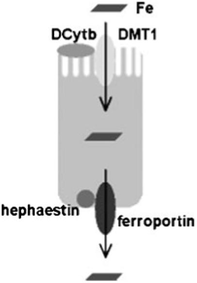
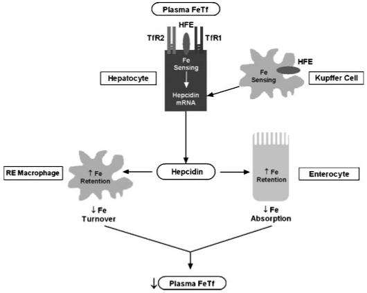
Hereditary hemochromatosis (HH) encompasses several inherited disorders of iron homeostasis characterized by increased gastrointestinal iron absorption and tissue iron deposition. The most common form of this disorder is HFE-related HH, nearly always caused by homozygosity for the C282Y mutation. A substantial proportion of C282Y homozygotes do not develop clinically significant iron overload, suggesting roles for environmental factors and modifier genes in determining the phenotype. Recent studies have demonstrated that the pathogenesis of nearly all forms of HH involves inappropriately decreased expression of the iron-regulatory hormone hepcidin. Hepcidin serves to decrease the export of iron from reticuloendothelial cells and absorptive enterocytes. Thus, HH patients demonstrate increased iron release from these cell types, elevated circulating iron, and iron deposition in vulnerable tissues. The mechanism by which HFE influences hepcidin expression is an area of current investigation and may offer insights into the phenotypic variability observed in persons with mutations in HFE.


Figures

Figure 1
Figure 1
Iron absorption by duodenal enterocytes. Duodenal villus cells are the major site of iron absorption from the diet. Prior to uptake, dietary ionic iron requires reduction from the ferric to the ferrous state. This is accomplished by ferric reductase(s) (for example, Dcytb) expressed on the luminal surface of villus enterocytes. Ferrous iron is taken up by the apical transporter, DMT1. Iron may be stored within the cell as ferritin and lost with the sloughed senescent enterocyte or transferred across the basolateral membrane to the plasma. This latter process occurs via the transporter ferroportin and is facilitated by oxidation of iron to the ferric state by the molecule hephaestin. Dcytb, duodenal cytochrome b related ferric reductase; DMT1, divalent metal transporter 1.
Figure 2
Figure 2
Role of hepcidin in the pathogenesis of hereditary hemochromatosis. In this model, diferric Tf in the portal circulation serves as the ligand for hepatocyte TfRs (TfR1 and/or TfR2). This interaction transduces a signal that increases hepatocellular expression of hepcidin. Alternatively, Kupffer cells may sense iron stores and send an as yet unknown signal to the hepatocytes to increase hepcidin expression. Circulating hepcidin binds ferroportin in target cells (duodenal enterocytes and reticuloendothelial macrophages) to decrease ferroportin-mediated iron export from these cells. In duodenal enterocytes, this leads to a decrease in the amount of dietary iron absorbed into the circulation. Thus, circulating levels of diferric Tf are normalized and homeostasis is maintained. In HFE-related HH, loss of functional HFE protein (in hepatocytes and/or Kupffer cells) leads to aberrant hepatocellular sensing of plasma iron, inappropriately low levels of hepcidin, decreased reticuloendothelial iron stores, and increased duodenal iron absorption. Tf, transferrin; TfR1, transferrin receptor 1; TfR2, transferrin receptor 2.

References

    1. Pietrangelo A. Hereditary hemochromatosis—a new look at an old disease. N Engl J Med. 2004;350:2383–2397. - PubMed
    1. Feder JN, Gnirke A, Thomas W, et al. A novel MHC class I-like gene is mutated in patients with hereditary haemochromatosis. Nat Genet. 1996;13:399–408. - PubMed
    1. Simon M, Bourel M, Fauchet R, et al. Association of HLA-A3 and HLA-B14 antigens with idiopathic haemochromatosis. Gut. 1976;17:332–334. - PMC - PubMed
    1. Zhou XY, Tomatsu S, Fleming RE, et al. HFE gene knockout produces mouse model of hereditary hemochromatosis. Proc Natl Acad Sci USA. 1998;95:2492–2497. - PMC - PubMed
    1. Bahram S, Gilfillan S, Kuhn LC, et al. Experimental hemochromatosis due to MHC class I HFE deficiency: immune status and iron metabolism. Proc Natl Acad Sci USA. 1999;96:13312–13317. - PMC - PubMed

Publication types

MeSH terms