. 2006 Aug;27(8):662-77.
doi: 10.1002/hbm.20209.

Attentional modulation of emotional stimulus processing: an fMRI study using emotional expectancy

Affiliations

Attentional modulation of emotional stimulus processing: an fMRI study using emotional expectancy

Felix Bermpohl et al. Hum Brain Mapp. 2006 Aug.

Abstract

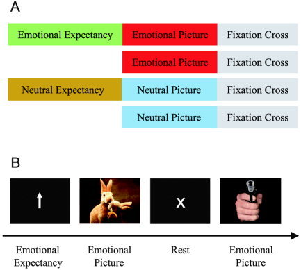
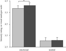
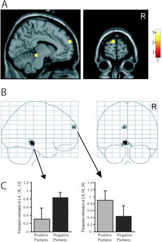
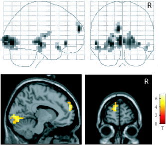
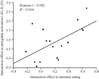
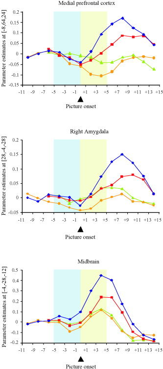
We used emotional expectancy to study attentional modulation in the processing of emotional stimuli. During functional magnetic resonance imaging (fMRI), volunteers saw emotional and neutral expectancy cues signaling the subsequent presentation of corresponding emotional or neutral pictorial stimuli. As a control, emotional and neutral pictures were presented without preceding expectancy cue, resulting in a 2 x 2 factorial design with the factors "expectancy" and "emotion." Statistical analysis revealed a significant positive interaction effect between these factors in the medial prefrontal cortex (MPFC, Brodmann area [BA] 9/10), amygdala, and dorsal midbrain. In all these regions, expectancy augmented the neural response to emotional but not to neutral pictures. Time course analysis of raw data suggests that this augmented activation was not preceded by baseline increases in MPFC and amygdala during the period of emotional expectancy. In a post-scanning session, the paradigm was presented for a second time to allow emotional intensity rating. Again, a significant interaction between expectancy and emotion was observed, with intensity ratings specifically enhanced in emotional photographs preceded by expectancy. There was a positive correlation between intensity ratings and blood oxygenation level-dependent (BOLD) signals in the left amygdala. We conclude that specific components of the emotion network show enhanced activation in response to emotional stimuli when these are preceded by expectancy. This enhancement effect is not present in neutral pictures and might parallel accentuated subjective feeling states.


Figures

Figure 1
Figure 1
Paradigm for fMRI. A: Experimental conditions. Participants viewed emotional and neutral photographs. In half of the trials these were preceded by an expectancy period indicating whether the subsequent picture was emotional or neutral. Each picture was followed by a fixation cross. B: Example trials. The trials “emotional picture with preceding expectancy period” and “emotional picture without preceding expectancy period” are illustrated. Stimuli are not drawn to scale.
Figure 2
Figure 2
Intensity rating. Emotional intensity of pictures was assessed using a nine‐point visual analogue scale. Larger values on the ordinate reflect higher intensity scores. Emotional pictures were rated significantly more intense when they were preceded by expectancy. Error bars show the standard error of the mean (SEM). *P < 0.05.
Figure 3
Figure 3
Positive interaction effect. Regions showing activation in the contrast (emotional picture with expectancy > emotional picture without expectancy) > (neutral picture with expectancy > neutral picture without expectancy), i.e., positive interaction between emotion and expectancy, presented in section A (through x = −8, y = 64, z = 24) and B glass‐brain views. P < 0.001 uncorrected. The bar diagram (C) separately indicates the contribution of positive (i.e., pleasant) and negative (i.e., unpleasant) picture conditions to the effects observed in midbrain and medial prefrontal cortex.
Figure 4
Figure 4
Emotional expectancy effect. Comparison between emotional conditions with and without preceding expectancy, presented in glass‐brain and section (through x = −8, y = 60, z = 20) views. The section views do not display all regions activated in this contrast (cf. Table II). P < 0.05 false discovery rate (FDR)‐corrected.
Figure 5
Figure 5
Correlation of interaction effects. Correlation between positive interaction effects in fMRI activation (left amygdala) and intensity ratings. The interaction effect on left amygdala activation was extracted from the peak voxel as identified by the positive interaction contrast (x = −20, y = −8, z = −20, cf. Table I). The interaction effect in intensity rating was determined by subtracting the expectancy effect in neutral pictures from the effect in emotional pictures.
Figure 6
Figure 6
Time histogram. Size of effect in the medial prefrontal cortex (MPFC), right amygdala, and dorsal midbrain over time. The curves represent the four conditions, emotional pictures with expectancy (rhomb), emotional pictures without expectancy (squares), neutral pictures with expectancy (circles), and neutral pictures without expectancy (triangles). The picture period (shaded area after picture onset) is preceded by the expectancy period (shaded area before picture onset) in the conditions with expectancy. In the conditions without expectancy, the picture presentation is preceded by the rest period. Values refer to the peak voxels as identified by the positive interaction contrast (cf. Table I): MPFC: x = −8, y = 64, z = 24. Right amygdala: x = 28, y = −4, z = −28. Dorsal midbrain: x = −4, y = −28, z = −12. The peak voxel in the left amygdala (x = −20, y = −8, z = −20) showed a time course similar to the right amygdala. Comparable findings were obtained when the analysis referred to clusters of activation (cf. Fig. 3A,B) instead of peak voxels.

References

    1. Alsop D (1995): Correction of ghost artifacts and distortion in echo‐planar MR imaging with an iterative image reconstruction technique. Radiology 197: 388.
    1. Amaral DG, Price JL, Pitkanen A, Carmichael ST. 1992. Anatomical organisation of the primate amygdaloid complex In: Aggleton JP, editor. The amygdala: neurobiological aspects of emotion, memory and mental dysfunction. New York: Wiley‐Liss; p 1–66.
    1. Anderson AK, Christoff K, Panitz D, De Rosa E, Gabrieli JD (2003a): Neural correlates of the automatic processing of threat facial signals. J Neurosci 23: 5627–5633. - PMC - PubMed
    1. Anderson AK, Christoff K, Stappen I, Panitz D, Ghahremani DG, Glover G, Gabrieli JD, Sobel N (2003b): Dissociated neural representations of intensity and valence in human olfaction. Nat Neurosci 6: 196–202. - PubMed
    1. Beauregard M, Levesque J, Bourgouin P (2001): Neural correlates of conscious self‐regulation of emotion. J Neurosci 21: RC165. - PMC - PubMed

Publication types

MeSH terms