

Anatomical changes in the emerging adult brain: a voxel-based morphometry study
- PMID: 16317714
- PMCID: PMC6871409
- DOI: 10.1002/hbm.20218
Anatomical changes in the emerging adult brain: a voxel-based morphometry study
Abstract
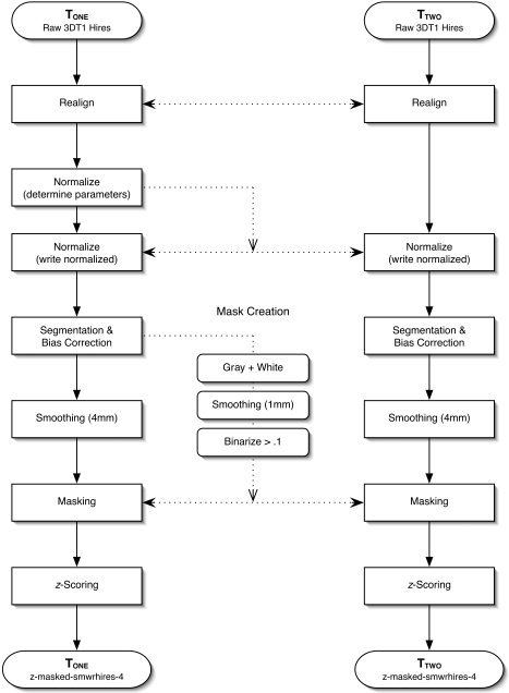
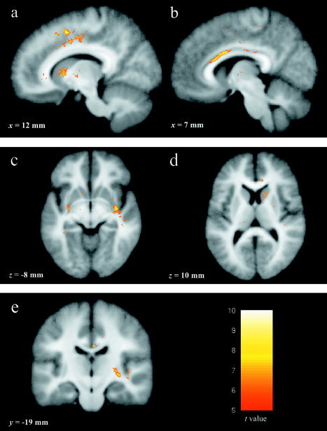
Research has consistently confirmed changes occur in brain morphometry between adolescence and adulthood. The purpose of the present study was to explore anatomical change during a specific environmental transition. High-resolution T1-weighted structural magnetic resonance imaging (MRI) scans were acquired from 19 participants (mean age at initial scan = 18.6 years) during their freshman year. Scans were completed during the fall term and 6 months later before the conclusion of the school year. Voxel-based morphometry was used to assess within-subject change. Significant intensity increases were observed along the right midcingulate, inferior anterior cingulate gyrus, right caudate head, right posterior insula, and bilateral claustrum. Regional changes were not observed in two control groups; one controlling for method and another controlling for age-specific change over time. The results suggest that significant age-related changes in brain structure continue after the age of 18 and may represent dynamic changes related to new environmental challenges. Findings from the regions of change are discussed in the context of specific environmental demands during a period of normative maturation.
(c) 2006 Wiley-Liss, Inc.
Figures
References
-
- Adolphs R (2003): Cognitive neuroscience of human social behaviour. Nat Rev Neurosci 4: 165–178. - PubMed
-
- Afifi AK (2003): The basal ganglia: a neural network with more than motor function. Semin Pediatr Neurol 10: 3–10. - PubMed
-
- Arnett JJ (2000): Emerging adulthood. A theory of development from the late teens through the twenties. Am Psychol 55: 469–480. - PubMed
-
- Arnett JJ (2004): Emerging adulthood: the winding road from the late teens through the twenties. New York: Oxford: Oxford University Press; 270 p.
-
- Ashburner J (2002): Another MRI bias correction approach. The 8th International Conference on Functional Mapping of the Human Brain, June 2–6, 2002, Sendai, Japan.
Publication types
MeSH terms
Grants and funding
LinkOut - more resources
Full Text Sources
Medical
