. 2006 Feb;17(2):895-906.
doi: 10.1091/mbc.e05-06-0512. Epub 2005 Nov 30.

Unusual kinetic and structural properties control rapid assembly and turnover of actin in the parasite Toxoplasma gondii

Affiliations

Unusual kinetic and structural properties control rapid assembly and turnover of actin in the parasite Toxoplasma gondii

Nivedita Sahoo et al. Mol Biol Cell. 2006 Feb.

Abstract

Toxoplasma is a protozoan parasite in the phylum Apicomplexa, which contains a number of medically important parasites that rely on a highly unusual form of motility termed gliding to actively penetrate their host cells. Parasite actin filaments regulate gliding motility, yet paradoxically filamentous actin is rarely detected in these parasites. To investigate the kinetics of this unusual parasite actin, we expressed TgACT1 in baculovirus and purified it to homogeneity. Biochemical analysis showed that Toxoplasma actin (TgACT1) rapidly polymerized into filaments at a critical concentration that was 3-4-fold lower than conventional actins, yet it failed to copolymerize with mammalian actin. Electron microscopic analysis revealed that TgACT1 filaments were 10 times shorter and less stable than rabbit actin. Phylogenetic comparison of actins revealed a limited number of apicomplexan-specific residues that likely govern the unusual behavior of parasite actin. Molecular modeling identified several key alterations that affect interactions between monomers and that are predicted to destabilize filaments. Our findings suggest that conserved molecular differences in parasite actin favor rapid cycles of assembly and disassembly that govern the unusual form of gliding motility utilized by apicomplexans.


Figures

Figure 1.
Figure 1.
Expression, purification, and in vitro polymerization of His6-TgACT1. (A) Phase-contrast and immunofluorescence images of His6-TgACT1 expression in SF9 cells. Stained with rabbit α-TgACT1 followed by secondary antibody conjugated to Alexa 488. Scale, 20 μm. (B) TgACT1 expressing SF9 (lane 1) and control SF9 (lane 2) cell lysates resolved by 10% SDS-PAGE and Western-blotted with anti-His tag antibody (α-His). Ni-resin chromatography purified TgACT1 (lane 3) resolved on 10% SDS-PAGE and stained with SYPRO Ruby. (C) Separation of polymerized and soluble His6-TgACT1 (0.5 μM) after addition of salts to G-buffer. After ultracentrifugation, the supernatants (s) and pellets (p) were analyzed on 10% SDS-PAGE and stained by SYPRO Ruby. (D) Different concentrations of His-TgACT1 were polymerized in F-buffer, and the ultracentrifuged6samples were analyzed by SDS-PAGE followed by Western blotting with anti-actin mAb C4 and peroxidase-conjugated goat anti-mouse IgG. The pellet fractions were quantified by phosphorimager analysis and plotted as relative light units (Y-axis). Actin polymerization occurred linearly above 0.03 μM (X-intercept). Curve fit using linear regression analysis.
Figure 2.
Figure 2.
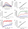
Polymerization properties of TgACT1. (A) Tryptophan quenching curves are shown for various concentrations (in nM) of untagged TgACT1 in G- or F-buffer. Readings were conducted 15 min after addition of F-buffer salts. Y-axis values are arbitrary fluorescence units. (B) Plotting the change in peak fluorescence between G-buffer and F-buffer (shown in A) versus concentration of untagged TgACT1, reveals the critical concentration to be ∼0.04 μM (X-intercept). (C) Plot of the change in tryptophan quenching versus concentration of TgACT1 in the presence of 10 μM CytD. The resulting Cc was ∼0.18 μM (X-intercept). (D) Polymerization of different concentrations of TgACT1 in 1.0 mM ATP and 2 mM MgCl2 over time as determined by tryptophan quenching. At low concentrations, a noticeable lag is observed. (E) Dependence of polymerization on ATP versus ADP. Curves show tryptophan quenching over time for 5.0 μM TgACT1. (F) Determination of the TgACT1 Cc in the presence of 1.0 mM ADP as determined by tryptophan quenching. The resulting Cc was ∼0.16 μM (X-intercept). The plots in B-F show the absolute value of the change in intrinsic fluorescence on the Y-axis in arbitrary fluorescence units.
Figure 3.
Figure 3.
In vitro polymerization of actin detected by epifluorescence microscopy. (A) Polymerization of rabbit actin (RbActin) alone leads to a large network of intertwined filaments revealed by staining with Alexa 488-phalloidin. (B) In contrast, polymerization of TgACT1 leads to formation of small foci that stain weakly with phalloidin but that react to rabbit anti-TgACT1 antibodies (revealed by staining with Alexa 594-conjugated goat anti-rabbit IgG). (C) Polymerization of a 1:1 mixture of these two proteins together did not result in copolymerization. Scale bars, 5 μm.
Figure 4.
Figure 4.
Ultrastructural examination of polymerized TgACT1 filaments compared with rabbit actin. (A) Filaments formed by TgACT1 were short, unbranched, and often appeared bent or broken (TgACT1 control). When polymerized TgACT1 filaments were mixed with 5 μM phalloidin on the grid, they were straight and had more distinct edges, indicating they were stabilized. TgACT1 filaments were observed to form pairs or small bundles when polymerized in the presence of JAS (1 μM; see bottom panel for enlargement). Rabbit actin formed long straight filaments with a characteristic helical spiral. Enlarged views (bottom panels) demonstrate the repeat striations, helical pattern, and ∼9-nm diameter of the filaments formed by TgACT1 and rabbit actin. After polymerization of 5 μM actins in F-buffer, 100,000 × g pellets were resuspended in 1× F-buffer, negatively stained with uranyl acetate, and examined by EM. (B) Histogram showing the distribution of filament lengths between control, phalloidin (5 μM) or JAS (1 μM) treated TgACT1 and rabbit muscle actin. TgACT1 filaments were more than 10-fold shorter than rabbit actin filaments formed under similar conditions. Treatment of TgACT1 with phalloidin or JAS increased the average size.
Figure 5.
Figure 5.
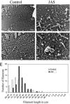
Visualization of actin filaments formed in parasites. Actin filaments were observed on the inner surface of the parasite plasma membrane that remained attached to the substrate after removal of the parasite by gentle sonication. Images were generated by rapid freeze-drying and platinum-coated replicas that were examined by EM. Scale bar, 100 nm in all panels. (A) In control cells, short filament arrays were detected on the inner surface of the parasite plasma membrane. (B) Enlargement of image shown in A showing the parallel arrangement of filaments. (C) Treatment with JAS (1 μM) resulted in a tangled web of short filaments. Most filaments were twice the normal diameter indicating they were bundled. Arrows denote single filaments. (D) Enlargement of a preparation similar to C showing individual TgACT1 filaments formed in the presence of JAS (arrows). (E) Average lengths of TgACT1 filaments formed in control (□) versus JAS-treated (▪) parasites, JAS treatment resulted in a slight increase in average length.
Figure 6.
Figure 6.
Phylogenetic and modeling analyses reveal conserved residues in parasite actins. (A) Phylogenetic analysis depicts the similarity of parasite actins to ciliates and dinoflagellates. The shaded circles show confidence estimates for trees (N = 1000 permutations). Sequence accession numbers for each actin are given in Materials and Methods. (B) Model of TgACT1 structure depicting apicomplexan specific residues. Amino acids conserved among various organisms are projected on to an ADP actin structure based on their sequence alignment. Color code refers to conservation of residues as follow: red, conserved in most apicomplexans (typically all except Cryptosporidium); orange, conserved in all apicomplexans; yellow, conserved in apicomplexans, ciliates, and/or dinoflagellates.
Figure 7.
Figure 7.
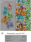
Conserved features of apicomplexan actins that are predicted to destabilize filaments. (A) Actin filament models were based on the Holmes structure. Molecular modeling was used to determine contact points between adjacent monomers. The hydrophobic plug is shown in yellow. (B) Salt bridge between R39 and E276 of adjacent monomers in the filament formed by muscle actin. Within the hydrophobic plug (yellow) the position 269 is M. (C) In TgACT1, the alteration of residue 277 in subdomain 3 from E to R eliminates the potential salt bridge with R40 in subdomain 2, resulting instead in charge repulsion between adjacent monomers in the filament. Within the hydrophobic plug (yellow), residue 270 is changed from M to K: the resulting positive charge is likely to alter interaction with the hydrophobic pocket in the adjacent monomer. Both of these changes have the potential to destabilize the filament. (D) Residues of the hydrophobic plug for muscle, yeast, and TgACT1 showing the hydrophobic nature of this region that is disrupted by alteration of M/L to K in T. gondii.

References

    1. Allen, M. L., Dobrowolski, J. M., Muller, H., Sibley, L. D., and Mansour, T. E. (1997). Cloning and characterization of actin depolymerizing factor from Toxoplasma gondii. Mol. Biochem. Parasitol. 88, 43-52. - PubMed
    1. Baldauf, S. L., Roger, A. J., Wenk-Siefert, I., and Doolittle, W. F. (2000). A kingdom-level phylogeny of eukaryotes based on combined protein data. Science 290, 972-977. - PubMed
    1. Barragan, A., and Sibley, L. D. (2003). Migration of Toxoplasma gondii across biological barriers. Trends Microbiol. 11, 426-430. - PubMed
    1. Bubb, M. R., Senderowicz, A.M.J., Sausville, E. A., Duncan, K.L.K., and Korn, E. D. (1994). Jasplakinolide, a cytotoxic natural product, induces actin polymerization and competitively inhibits the binding of phalloidin to F-actin. J. Biol. Chem. 269, 14869-14871. - PubMed
    1. Bubb, M. R., Spector, I., Beyer, B. B., and Fosen, K. M. (2000). Effects of jasplakinolide on the kinetics of actin polymerization. J. Biol. Chem. 275, 5163-5170. - PubMed

Publication types

MeSH terms

LinkOut - more resources