. 2005 Nov 30;25(48):11061-70.
doi: 10.1523/JNEUROSCI.3034-05.2005.

Soluble beta-amyloid1-40 induces NMDA-dependent degradation of postsynaptic density-95 at glutamatergic synapses

Affiliations

Soluble beta-amyloid1-40 induces NMDA-dependent degradation of postsynaptic density-95 at glutamatergic synapses

F Roselli et al. J Neurosci. .

Abstract

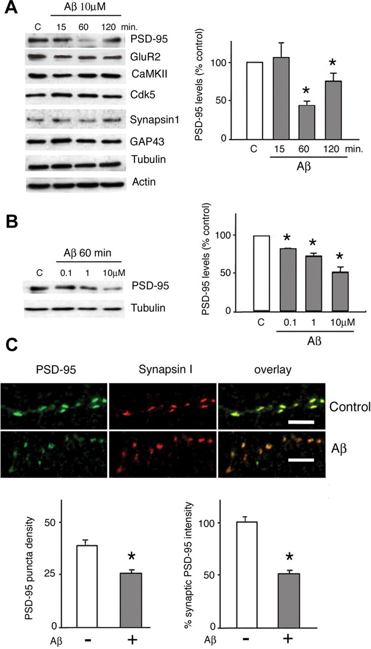
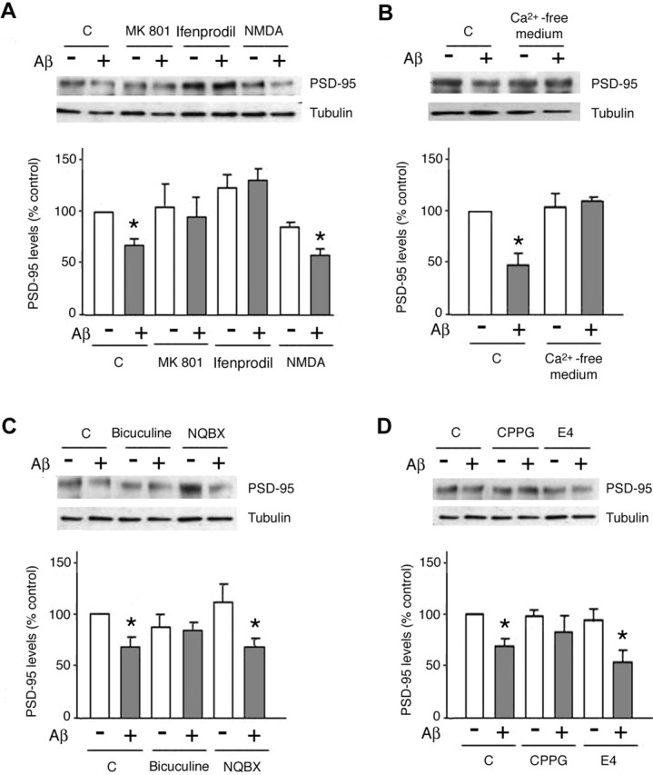
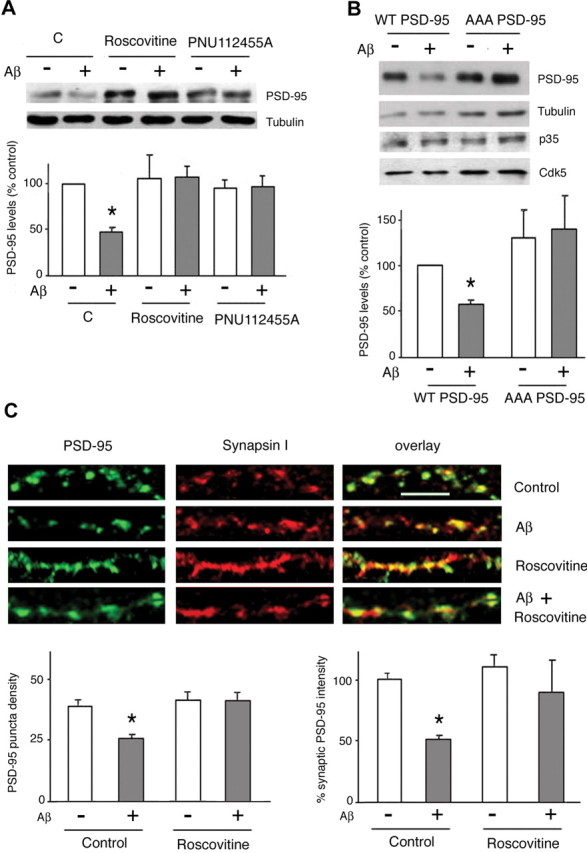
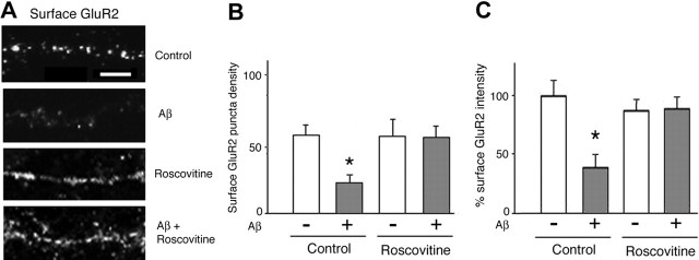
Amyloid-beta (Abeta) has been implicated in memory loss and disruption of synaptic plasticity observed in early-stage Alzheimer's disease. Recently, it has been shown that soluble Abeta oligomers target synapses in cultured rat hippocampal neurons, suggesting a direct role of Abeta in the regulation of synaptic structure and function. Postsynaptic density-95 (PSD-95) is a postsynaptic scaffolding protein that plays a critical role in synaptic plasticity and the stabilization of AMPA (AMPARs) and NMDA (NMDARs) receptors at synapses. Here, we show that exposure of cultured cortical neurons to soluble oligomers of Abeta(1-40) reduces PSD-95 protein levels in a dose- and time-dependent manner and that the Abeta1(1-40)-dependent decrease in PSD-95 requires NMDAR activity. We also show that the decrease in PSD-95 requires cyclin-dependent kinase 5 activity and involves the proteasome pathway. Immunostaining analysis of cortical cultured neurons revealed that Abeta treatment induces concomitant decreases in PSD-95 at synapses and in the surface expression of the AMPAR glutamate receptor subunit 2. Together, these data suggest a novel pathway by which Abeta triggers synaptic dysfunction, namely, by altering the molecular composition of glutamatergic synapses.


Figures

Figure 1.
Figure 1.
PSD-95 levels are reduced after Aβ treatment in rat primary cortical neurons. A, Aβ selectively downregulates PSD-95 levels in a time-dependent manner, without altering the expression of other synaptic proteins. Cells were exposed to 10 μm soluble Aβ1-40 (see Materials and Methods) for between 15 and 120 min before analysis by Western blot. As shown in the histogram, semiquantitative evaluation of PSD-95 levels, normalized against tubulin, shows that Aβ treatment led to a significant reduction of PSD-95 expression (53.3 ± 6.5% of control levels; p < 0.05) within 60 min; despite a trend to recover by 120 min after treatment, PSD-95 expression remained significantly lower than in controls (76 ± 10.9%; p < 0.05). Analysis of levels of the synaptic proteins GluR2, GAP-43, synapsin I, cdk5, and CaMKII revealed no significant effect of Aβ over the treatment duration. Actin and tubulin levels were not influenced by the experimental manipulations. B, Dose-dependent effects of Aβ on PSD-95 levels. Neurons were exposed to Aβ (0.1-10 μm) for 1 h before they were analyzed for levels of PSD-95 expression in Western blot assays. One representative Western blot is shown, and the semiquantitative data from three independent experiments are shown in the histogram. Levels of PSD-95, normalized with respect to tubulin levels, are shown as means ± SD. Asterisks indicate significant changes from untreated control cells (p < 0.05). The dose-response curve had r = 0.879 (p = 0.007). C, Aβ effect on synaptic PSD-95. Primary rat cortical neurons were treated with 10 μm Aβ for 1 h, fixed, and immunostained for synapsin I and PSD-95. Synaptic sites were identified as synapsin I-positive puncta. Puncta density in vehicle-treated control cultures was 36 ± 2.4 puncta/100 μm; after Aβ treatment, the density of PSD-95 immunoreactive puncta was 24.6 ± 2.1/100 μm (p < 0.001). Numerous synaptic sites (identified by synapsin staining) showed prominent decreases in PSD-95 fluorescence after Aβ treatment; fluorescence intensity of synaptic PSD-95 was reduced after Aβ treatment (49.4 ± 2.4% compared with 100 ± 6.7% in controls; p < 0.01, p < 0.05; n = 200). Scale bars, 5 μm. All numerical data represent mean ± SD.
Figure 2.
Figure 2.
Aβ-induced PSD-95 downregulation requires NMDAR activity and calcium influx. A, Aβ-induced decrease in PSD-95 levels requires NMDAR activity. Cells treated with Aβ showed a significant decrease in PSD-95 levels (67.5 ± 6.7%; p < 0.05 vs untreated cells). Treatment (1 h) of cells with MK-801 (10 μm) or ifenprodil (10 μm) prevented the Aβ-induced decrease in PSD-95 levels, whereas NMDA (10 μm) did not influence the effects of Aβ (59 ± 5%; p < 0.05 compared with nontreated cells). C, Control. B, PSD-95 downregulation by Aβ is a calcium-dependent process. Neurons exposed to Aβ (10 μm; 1 h) under calcium-free conditions did not show a reduction in PSD-95 levels compared with untreated control cells. C, The PSD-95 down-regulating actions of Aβ are not dependent on AMPAR and are attenuated by bicuculline. Neurons were pretreated (1 h) with NBQX (20 μm), bicuculline (40 μm), or vehicle before exposure to Aβ (10 μm; 1 h). NBQX proved ineffective in counteracting Aβ action (61.9 ± 9.1%), whereas bicuculline attenuated the effects of Aβ on PSD-95 levels (81.4 ± 15%; p < 0.05 vs Aβ alone). D, The Aβ-induced decrease in PSD-95 levels is blocked in the presence of a metabotropic II/III receptor antagonist. Neurons were pretreated (1 h) with the mGluRI/II antagonist E4CPG (E4) (10 μm), the mGluRII/III antagonist CPPG (10 μm), or vehicle before treatment with Aβ (10 μm) for an additional 1 h. E4CPG did not alter the actions of Aβ on PSD-95, whereas CPPG significantly antagonized the Aβ effect (81.4 ± 15%; p > 0.05 vs response to Aβ only). Asterisks indicate significant changes from untreated control cells (p < 0.05). All data are given as mean ± SD.
Figure 3.
Figure 3.
Aβ-induced downregulation of synaptic PSD-95 requires cdk5 activity. A, cdk5 activity is necessary for Aβ effects in cortical neurons. Primary rat cortical neurons were pretreated (1 h) with roscovitine (15 μm) or PNU 112455A (10 μm), or vehicle (DMSO) before exposure to Aβ (10 μm; 1 h). As shown by Western blotting, both inhibitors abrogated the ability of Aβ to reduce PSD-95 levels. C, Control. B, Aβ cannot downregulate PSD-95 levels when SK-N-MC cells are transiently transfected with the T19A, S25A, S35A phosphorylation mutant of PSD-95. Cells transfected with wild-type PSD-95 and expressing p35/cdk5 responded to Aβ with the expected decrease in PSD-95 levels (57.9 ± 5.6% of controls; p < 0.05). C, The Aβ effects on synaptic PSD-95 are blocked by roscovitine, an inhibitor of cdk5 activity. Primary rat cortical neurons were pretreated (1 h) with roscovitine (15 μm) or vehicle (DMSO) before exposure to Aβ (10 μm; 1 h). After fixation, neurons were immunostained for synapsin I and PSD-95. Roscovitine alone did not show a significant effect on the density of PSD-95 puncta or their fluorescence intensity but inhibited the effects of Aβ on these parameters. Scale bar, 5 μm. Asterisks indicate significant changes from untreated control cells (p < 0.05).
Figure 4.
Figure 4.
The proteasome pathway is implicated in the regulation of PSD-95 by Aβ. A, PSD-95-transfected cultures were pretreated (1 h) with a proteasome inhibitor (MG132; 0.1 μm), a lysosome inhibitor (chloroquine; 100 μm), or vehicle (DMSO) before treatment with Aβ (10 μm; 1 h). MG132 treatment prevented Aβ-induced reductions in PSD-95, whereas chloroquine did not exert a significant influence on the actions of Aβ. C, Control. B, The PEST consensus sequence in PSD-95 is necessary for the Aβ-induced effects. SK-N-MC cultures that had been transfected with either wild-type PSD-95 or the ΔPEST deletion mutant of PSD-95 were exposed to Aβ (10 μm; 1 h). Whereas Aβ led to a reduction in PSD-95 levels in wild-type transfected cells (53.6 ± 19.8%; p < 0.05 vs non-Aβ-treated controls), PSD-95 levels were unchanged in cells expressing the ΔPEST mutant. Asterisks indicate significant changes from untreated control cells (p < 0.05).
Figure 5.
Figure 5.
Aβ reduces the expression of surface AMPARs. Primary rat cortical neurons were immunostained for the AMPAR subunit of the GluR2; permeabilization steps were excluded to ensure labeling of surface receptors. A, B, The density of GluR2-positive puncta (expressed as the number of puncta per 100 μm) was reduced after Aβ treatment (21.2 ± 6.5 vs 55.5 ± 7.2; p < 0.001). A, C, Aβ treatment led to a significant reduction in the intensity of immunofluorescence of GluR2 puncta (38.6 ± 11.6%; p < 0.001; 225 puncta analyzed). Pretreatment with roscovitine (15 μm) abrogated the effects of Aβ on these parameters (p < 0.05) but had no effect of its own. Scale bar, 5 μm. Asterisks indicate significant changes from untreated control cells (p < 0.05).

References

    1. Bingol B, Schuman E (2004) A proteasome-sensitive connection between PSD-95 and GluR1 endocytosis. Neuropharmacology 47: 755-763. - PubMed
    1. Bitan G, Lomakin A, Teplow DB (2001) Amyloid β protein oligomerization prenucleation interactions revealed by photo-induced cross-linking of unmodified proteins. J Biol Chem 276: 35176-35184. - PubMed
    1. Bitan G, Vollers SS, Teplow DB (2003) Elucidation of primary structure elements controlling early amyloid beta-protein oligomerization. J Biol Chem 278: 34882-34889. - PubMed
    1. Bredt D, Nicoll R (2003) AMPA receptor trafficking at excitatory synapses. Neuron 40: 361-379. - PubMed
    1. Chen L, Chetkovich DM, Petralia RS, Sweeney NT, Kawasaki Y, Wenthold RJ, Bredt DS, Nicoll RA (2000) Stargazin regulates synaptic targeting of AMPA receptors by two distinct mechanisms. Nature 408: 936-943. - PubMed

Publication types

MeSH terms

LinkOut - more resources