. 2005 Dec 1;438(7068):633-8.
doi: 10.1038/nature04321.

Lipid-protein interactions in double-layered two-dimensional AQP0 crystals

Affiliations

Lipid-protein interactions in double-layered two-dimensional AQP0 crystals

Tamir Gonen et al. Nature. .

Erratum in

  • Nature. 2006 May 11;441(7090):248

Abstract

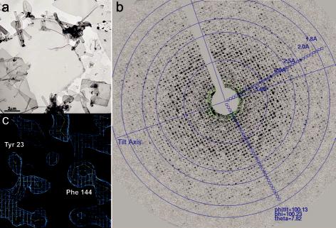
Lens-specific aquaporin-0 (AQP0) functions as a specific water pore and forms the thin junctions between fibre cells. Here we describe a 1.9 A resolution structure of junctional AQP0, determined by electron crystallography of double-layered two-dimensional crystals. Comparison of junctional and non-junctional AQP0 structures shows that junction formation depends on a conformational switch in an extracellular loop, which may result from cleavage of the cytoplasmic amino and carboxy termini. In the centre of the water pathway, the closed pore in junctional AQP0 retains only three water molecules, which are too widely spaced to form hydrogen bonds with each other. Packing interactions between AQP0 tetramers in the crystalline array are mediated by lipid molecules, which assume preferred conformations. We were therefore able to build an atomic model for the lipid bilayer surrounding the AQP0 tetramers, and we describe lipid-protein interactions.


Figures

Figure 1
Figure 1
: Electron crystallography of AQP0 junctions. a. Double-layered AQP0 2D crystals were often several μm in size. b. A typical electron diffraction pattern recorded from an untilted AQP0 2D crystal prepared by the carbon sandwich technique showing diffraction spots to a resolution beyond 2 Å. c. Region of the final 2Fo-Fc map of AQP0 refined to 1.9 Å resolution. Two aromatic residues, Tyr 23 and Phe 144, that line the water pore in AQP0 are represented by donut-shaped densities.
Figure 2
Figure 2
: Structural differences between junctional and non-junctional AQP0. a. X-ray structure of non-junctional AQP0 (blue) superimposed on the EM structure of junctional AQP0 (yellow). The arrow indicates the conformational switch of extracellular loop A. b. Interactions involving the C-terminus of full-length AQP0. c. Interactions involving the N-terminus of full-length AQP0. d. C-terminus of cleaved AQP0. e. N-terminus of cleaved AQP0. f. View of the extracellular surface of the X-ray structure of non-junctional, full-length AQP0. g. View of the extracellular surface of the EM structure of junctional AQP0 from the lens core, showing the rosette-like structure formed by the Pro 38 residues.
Figure 3
Figure 3
: The water pore in AQP0. a. The pore in non-junctional AQP0 (left) contains seven water molecules (red spheres), while the pore in junctional AQP0 contains only three water molecules (right). Calculated pore profiles (middle) corroborate that the pore in junctional AQP0 (purple) is more constricted than in non-junctional AQP0 (pink). b. Hydrogen bonding pattern of water molecules in the pore of non-junctional AQP0 (left) and junctional AQP0 (right). The hydrogen bonding network is disrupted by Tyr 24, which introduces a phenolic barrier. In junctional AQP0 all three water molecules are too far apart to form hydrogen bonds. Dotted lines represent hydrogen bonds. See Supplementary Figure 5.
Figure 4
Figure 4
: Lipid-protein interactions in double-layered AQP0 2D crystals. a. Vertical slab through the 2Fo-Fc density map with modelled lipid molecules, revealing the two lipid bilayers in the double-layered AQP0 2D crystal. b. The nine lipids surrounding an AQP0 monomer in the 2D crystal. Lipids PC1 to PC7 are annular lipids, whereas lipids PC8 and PC9 are bulk lipids with no direct protein contacts. See Supplementary Figure 6 for a stereo view. c – e. Three examples of lipids sandwiched in between two AQP0 molecules. The acyl chains of PC1 adopt a closed (c), those of PC5 a slightly splayed (d), and those of PC6 a widely splayed conformation (e).

Comment in

  • Cell biology: a greasy grip.
    Lee AG. Lee AG. Nature. 2005 Dec 1;438(7068):569-70. doi: 10.1038/438569a. Nature. 2005. PMID: 16319869 No abstract available.

References

    1. Agre P, et al. Aquaporin water channels--from atomic structure to clinical medicine. J. Physiol. 2002;542:3–16. - PMC - PubMed
    1. Murata K, et al. Structural determinants of water permeation through aquaporin-1. Nature. 2000;407:599–605. - PubMed
    1. Fu D, et al. Structure of a glycerol-conducting channel and the basis for its selectivity. Science. 2000;290:481–486. - PubMed
    1. Sui H, Han BG, Lee JK, Walian P, Jap BK. Structural basis of water-specific transport through the AQP1 water channel. Nature. 2001;414:872–878. - PubMed
    1. Ren G, Reddy VS, Cheng A, Melnyk P, Mitra AK. Visualization of a water-selective pore by electron crystallography in vitreous ice. Proc. Natl. Acad. Sci. U. S. A. 2001;98:1398–1403. - PMC - PubMed

Publication types