. 2005 Dec 21;24(24):4247-59.
doi: 10.1038/sj.emboj.7600886. Epub 2005 Dec 1.

Structure of the entire cytoplasmic portion of a sensor histidine-kinase protein

Affiliations

Structure of the entire cytoplasmic portion of a sensor histidine-kinase protein

Alberto Marina et al. EMBO J. .

Abstract

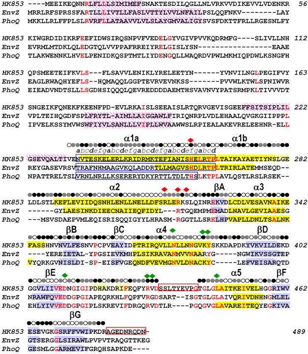
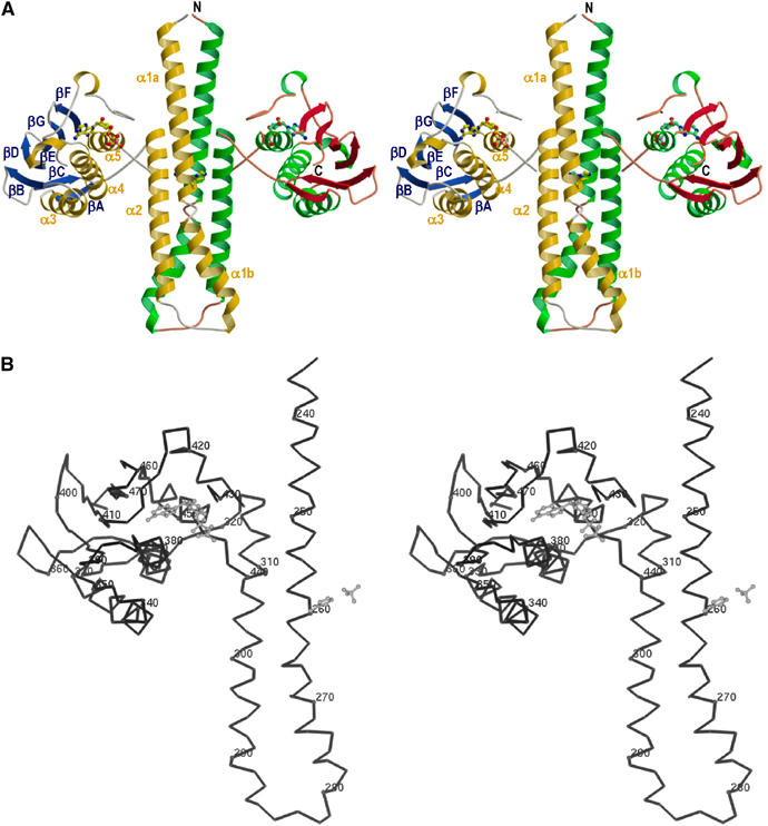
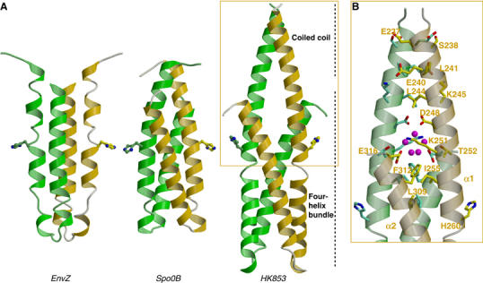
The large majority of histidine kinases (HKs) are multifunctional enzymes having autokinase, phosphotransfer and phosphatase activities, and most of these are transmembrane sensor proteins. Sensor HKs possess conserved cytoplasmic phosphorylation and ATP-binding kinase domains. The different enzymatic activities require participation by one or both of these domains, implying the need for different conformational states. The catalytic domains are linked to the membrane through a coiled-coil segment that sometimes includes other domains. We describe here the first crystal structure of the complete cytoplasmic region of a sensor HK, one from the thermophile Thermotoga maritima in complex with ADPbetaN at 1.9 A resolution. The structure reveals previously unidentified functions for several conserved residues and reveals the relative disposition of domains in a state seemingly poised for phosphotransfer. The structure thereby inspires hypotheses for the mechanisms of autophosphorylation, phosphotransfer and response-regulator dephosphorylation, and for signal transduction through the coiled-coil segment. Mutational tests support the functional relevance of interdomain contacts.


Figures

Figure 1
Figure 1
Sequence alignment of TM0853, EnvZ and PhoQ HKs. The three amino-acid sequences are aligned based on their structures. β sheets are shown as blue arrows and α helices as yellow filled boxes. Transmembrane regions predicted by the DAS program (Cserzo et al, 1997) are shown as purple intermediate shading and coiled-coil motifs predicted by LEARNCOIL program (Singh et al, 1998) are enclosed in blue boxes showing the helical position from a to g. Disordered regions are enclosed in red boxes. Residues identical in all three sequences are colored in red. The solvent accessibility of the HK853–CD is indicated for each residue by an open circle if the fraction solvent accessibility is >0.4, a half-filled circle if it is 0.1–0.4 and a filled circle if it is <0.1. Residues that interact with the ADPβN and the sulfate ion in HK853–CD are indicated by green and red diamonds, respectively.
Figure 2
Figure 2
In vitro and in vivo HK853 activity. (A) Time course of in vitro autophosphorylation of HK853–CD with [γ-32P]ATP. In all, 1–2 μM of HK853–CD was incubated in reaction buffer and samples were removed at indicated time points, reaction stopped by addition of SDS–PAGE sample buffer, subjected to gel electrophoresis, and phosphorylated protein was visualized by phosphorimaging. (B) Activation of the PhoQ–PhoP and EnvZ–OmpR HK–RR systems. E. coli reporter strains containing a PhoP-activated lacZ, but devoid of PhoQ (ΔPhoQ), or an OmpR-activated lacZ, but devoid of EnvZ (ΔEnvZ), were transformed with the pBR322-derived plasmids pHK853, pEnvZ, pPhoQ or pBR322 (expressing the respective full-length sensor kinases or a negative control). Response was assayed by β-galactosidase activity. The low activation by PhoQ was due to repressing divalent cations in culture media.
Figure 3
Figure 3
Molecular structure of the cytoplasmic portion of TM0853. (A) Ribbon representation of the crystallographic dimer of HK853–CD, including ADPβN. The α helices are labeled α1–α5, and colored gold (subunit A) and green (subunit B), β strands are labeled βA–βF, and colored blue (subunit A) and red (subunit B). The positions of N and C termini are labeled in subunit B. The ADPβN molecule and the phospho-acceptor residue (His260) are shown in ball-and-stick representation. The membrane would be located on the top of N-terminal residues. (B) Stereo Cα trace of the gold and blue protomer. Every tenth Cα is indicated as a sphere and numbered. The ADPβN molecule, the His260 and the sulfate ion coordinated with His260 are drawn in a gray ball-and-stick representation. The orientation is as in panel (A).
Figure 4
Figure 4
Comparison of DHp domains of EnvZ HK, Spo0B phosphotransferase and TM0853 HK. (A) Ribbon diagrams of the three DHp domains in their dimeric forms (protomers colored in green and gold). The DHp domains have been oriented with the plane containing the phospho-accepting histidines, which are shown in stick representation, and the principal helix axes parallel to the page. (B) Detail of the HK853 coiled-coil motif. Residues interacting in this motif are shown as stick representation and labeled on one protomer. Solvent molecules (magenta) are shown in the cavity generated at the juncture between the coiled coil of α1a helices and the four-helix bundle.
Figure 5
Figure 5
Comparison of the nucleotide-binding site of the TM0853 and PhoQ HKs. Secondary structures surrounding the ATP-binding site are drawn as gray ribbons and the ATP lids are in magenta. The nucleotides and the residues interacting with their phosphates are depicted as sticks and labeled. The Mg2+ ion of PhoQ is drawn as a cyan sphere. Hydrogen bonds are shown as dotted lines. Electron density of the HK853 nucleotide is contoured as a semitransparent blue surface at a level of 1σ.
Figure 6
Figure 6
Interactions between DHp and CA domains. Stereoview of the structural elements involved in interdomain contacts and sulfate ion interactions. The DHp domain, CA domain and interdomain-connecting loop are represented in blue, gold and green ribbon diagrams, respectively. Additionally, the α1′ helix, which presents His260′ as a sulfate ligand, is shown in gray. The interacting side chains are shown as sticks with the same carbon atom color as the corresponding domain, except the sulfate-interacting residues that are in gray. Nitrogen, oxygen, sulfur and nucleotide molecule are drawn in blue, red, black and magenta, respectively. Residue labels take the colors of their domains. Hydrogen bonds and salt bridges between the sulfate ion and interacting residues are indicated by purple dots.
Figure 7
Figure 7
Autokinase activity of interfacial mutant variants. (A) Mutations at the interfacial residue Ile448. (B) Double-cysteine mutations when reduced by DTT (open symbols) and when oxidized (filled symbols), where the negligible activity is overlapping. (C) Mutations to proline in the linker segment.
Figure 8
Figure 8
Structure-based schematic of the reactions catalyzed by HK sensors. The kinase autophosphorylation (A → B), phosphotransferase (B → A*) and phosphatase (A* → A) activities are shown on projected outlines of the enzyme and protein–substrate models. Positions of N and C termini, ATP and the phospho-accepting histidine (H) are indicated on an HK dimer (orange and green). Position of phospho-accepting aspartate (D) is indicated on a RR (red). The transferred phosphoryl group is indicated as a yellow asterisk.
Figure 9
Figure 9
Models of complexes for the phosphotransferase and kinase reactions catalyzed by HK853-CD. (A) Ribbon representation of the experimental complex (Zapf et al, 2000) between Spo0B (green and yellow) and Spo0F (red). For clarity, only one Spo0F molecule is drawn. (B) The Spo0F RR (red) docked onto the HK853–CD dimer (green and yellow) as a model of the phosphotransferase complex (see text). The catalytic histidine and aspartate residues and the nucleotide are shown as stick models. (C) Orthogonal view of (B). (D) Model of HK853–CD poised for the autokinase reaction. The catalytic domain of one protomer has been moved to align the γ phosphate of its ATP moiety with the phosphoaccepting histidine of the other promoter to permint trans-phosphorylation. The histidine and nucleotide are shown in stick representation. (E) Orthogonal view of (D).

References

    1. Aravind L, Ponting CP (1999) The cytoplasmic helical linker domain of receptor histidine kinase and methyl-accepting proteins is common to many prokaryotic signalling proteins. FEMS Microbiol Lett 176: 111–116 - PubMed
    1. Bilwes AM, Alex LA, Crane BR, Simon MI (1999) Structure of CheA, a signal-transducing histidine kinase. Cell 96: 131–141 - PubMed
    1. Bilwes AM, Quezada CM, Croal LR, Crane BR, Simon MI (2001) Nucleotide binding by the histidine kinase CheA. Nat Struct Biol 8: 353–360 - PubMed
    1. Birck C, Mourey L, Gouet P, Fabry B, Schumacher J, Rousseau P, Kahn D, Samama JP (1999) Conformational changes induced by phosphorylation of the FixJ receiver domain. Struct Fold Des 7: 1505–1515 - PubMed
    1. Butler SL, Falke JJ (1998) Cysteine and disulfite scanning reveals two amphiphilic helices in the linker region of the aspartate chemoreceptor. Biochemistry 37: 10746–10756 - PMC - PubMed

Publication types

MeSH terms