. 2006 Jan 10;494(2):358-67.
doi: 10.1002/cne.20814.

Cytoskeletal organization of the developing mouse olfactory nerve layer

Affiliations

Cytoskeletal organization of the developing mouse olfactory nerve layer

Michael R Akins et al. J Comp Neurol. .

Abstract

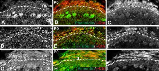
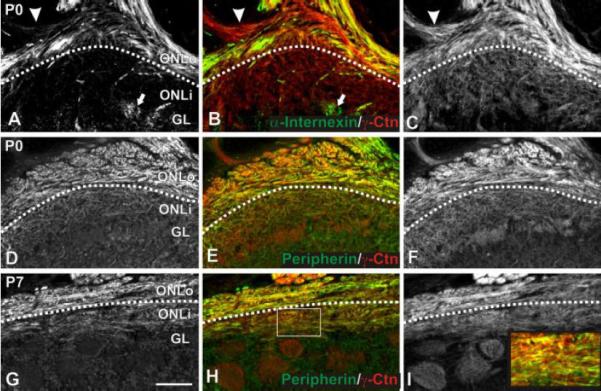
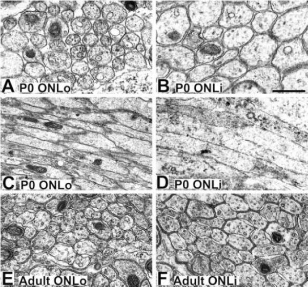
Olfactory sensory neuron (OSN) axonal extension and targeting occur within the olfactory nerve layer (ONL) of the olfactory bulb (OB). The ONL can be differentiated into sublaminae: the outer (ONLo), where axons broadly target regions of the OB in tight fascicles, and inner (ONLi), where axons perform final targeting in loosely organized fascicles. During perinatal development, cadherin-2 and its binding partner, gamma-catenin, are preferentially expressed by OSN axons in the ONLo vs. the ONLi. Given the expression of these cytoskeleton-associated molecules, we hypothesized that cytoskeletal elements of OSN axons may be differentially expressed across the ONL. We therefore examined cytoskeletal organization of OSN axons in the ONL, focusing on the day of birth (P0). We show that microfilaments, microtubules, and the intermediate filament (IF) vimentin are homogeneously expressed across the ONL at P0. In contrast, the IFs peripherin and alpha-internexin are preferentially localized to the ONLo at P0, with alpha-internexin expressed by a restricted subset of OSNs. We also show that OSN axons in the ONLo are significantly smaller than those in the ONLi. The data demonstrate that, as OSN axons begin to exit the ONLo and target a specific region of the OB, there is a down-regulation of cytoskeletal elements and bound extracellular adhesion molecules. The increase in axon diameter may reflect additional mechanisms involved in glomerular targeting or the formation of the large terminal boutons of OSN axons within glomeruli.


Figures

Figure 1
Figure 1
Localization of microfilaments, microtubules, and vimentin in the P0 ONL. γ-Catenin expression was enriched in the ONLo, allowing identification of the border between ONLo and ONLi (indicated by dotted line). (A-C) F-actin localization. Coronal sections were stained for phalloidin (green; A,B) and immunostained for γ-catenin (red; B,C). F-actin was seen uniformly across the ONL and most strongly in glomeruli. (D-F) Microtubule localization. Coronal sections were immunostained for α-tubulin (green; D,E) and γ-catenin (red; E,F). α-Tubulin was present ubiquitously within the OB. No difference in expression was seen between the ONLo and ONLi. (G-I) Vimentin localization. Coronal sections were immunostained for vimentin (green; G,H) and γ-catenin (red; H,I). Vimentin was expressed by OSN axons as well as non-neuronal tissue (e.g., blood vessel indicated by arrow in G,H). Within OSN axons, expression was uniform across the ONL. Scale bar = 100 μm. Abbreviations: ONLo, outer olfactory nerve layer; ONLi, inner olfactory nerve layer; Ctn, catenin; OSN, olfactory sensory neuron.
Figure 2
Figure 2
Localization of α-internexin and peripherin. γ-Catenin expression was used to identify the border between ONLo and ONLi as in Figure 1 for panels A-F. Peripherin expression allowed this distinction in G-I. (A-C) α-Internexin localization. Sagittal P0 sections were immunostained for α-internexin (green; A,B) and γ-catenin (red; B,C). α-Internexin was expressed by OSN axons within the ventral ONLo and dendrites within glomeruli in all regions of the OB (arrow in A,B). Expression was seen in only a subset of fascicles within the ONL (e.g., the fascicle indicated by an arrowhead in A,B is negative for α-internexin). (D-I) Peripherin localization. P0 (D-F) and P7 (G-I) coronal sections were immunostained for peripherin (green; D,E,G,H) and γ-catenin (red; E,F,H,I). At P0 (D-F), peripherin was found within OSN axons within the ONLo. Expression was observed in all fascicles. By P7 (G-I), peripherin expression had spread to the ONLi, although staining was more intense within the ONLo. Within the ONLi, staining was heterogeneous (box in H magnified as inset in I). Scale bar = 100 μm. Abbreviations: ONLo, outer olfactory nerve layer; ONLi, inner olfactory nerve layer; Ctn, catenin; OSN, olfactory sensory neuron.
Figure 3
Figure 3
Localization of cells expressing α-internexin. P0 sections were treated with a probe specific for α-internexin. (A) Expression was found in dorsolateral and ventromedial OE as well as in the SO (arrow). Expression is indicated with black lines in the right half of the section. (B) An enlargement of the dorsolateral region indicated in A. A sharp border of α-internexin expression was seen (indicated by the arrows). (C) An enlargement of the region indicated in B. α-Internexin was expressed by a subset of cells within the basal portion of the OE. (D) An expansion of the SO as indicated in A. Expression was seen in a subset of cells in the basal portion of the epithelium. (E) Expression within the VNO. α-Internexin was observed by a subset of cells located basally within the epithelium. Scale bar (shown in B) = 500 μm in A; 100 μm in B; 50 μm in C, D, E. Abbreviations: OE, olfactory epithelium; SO, septal organ; VNO, vomeronasal organ.
Figure 4
Figure 4
Summary of α-internexin expression. In situ hybridization of coronal P0 sections with a probe specific for α-internexin. Hemisections approximately every 100 μm are summarized in this figure. Presence of expression is indicated by a red line. Expression was seen within the VNO and SO. Additionally, expression was seen within the ventromedial and dorsolateral OE. Caudally, expression was seen ventrally. Scale bar = 1 mm. Abbreviations: OE, olfactory epithelium; SO, septal organ; VNO, vomeronasal organ.
Figure 5
Figure 5
Electron micrographs of axons in the P0 and adult ONL. (A,C) Axons within the P0 ONLo are of roughly uniform diameter. (B,D) Axons within the P0 ONLi are larger than those seen in the P0 ONLo. (E) Axons within the adult ONLo are approximately the same size seen in the P0 ONLo. (F) Axons within the adult ONLi are larger than those in the adult ONLo, but smaller than those in the P0 ONLi. Scale bar = 500 nm in A,B,E,F; 1 μm in C,D. Abbreviations: ONLo, outer olfactory nerve layer; ONLi, inner olfactory nerve layer.

References

    1. Akins MR, Greer CA. Catenins define axon paths in the developing mouse olfactory system. 2005. In submission.
    1. Ameen NA, Figueroa Y, Salas PJ. Anomalous apical plasma membrane phenotype in CK8-deficient mice indicates a novel role for intermediate filaments in the polarization of simple epithelia. J Cell Sci. 2001;114:563–575. - PubMed
    1. Astic L, Saucier D. Anatomical mapping of the neuroepithelial projection to the olfactory bulb in the rat. Brain Res Bull. 1986;16:445–454. - PubMed
    1. Astic L, Saucier D. Topographical projection of the septal organ to the main olfactory bulb in rats: ontogenetic study. Brain Res. 1988;470:297–303. - PubMed
    1. Astic L, Saucier D, Holley A. Topographical relationships between olfactory receptor cells and glomerular foci in the rat olfactory bulb. Brain Res. 1987;424:144–152. - PubMed

Publication types

MeSH terms