. 2005 Dec;52(12):4018-27.
doi: 10.1002/art.21485.

Identification of polyclonal and monoclonal antibodies against tissue plasminogen activator in the antiphospholipid syndrome

Affiliations

Identification of polyclonal and monoclonal antibodies against tissue plasminogen activator in the antiphospholipid syndrome

Cai-Sheng Lu et al. Arthritis Rheum. 2005 Dec.

Abstract

Objective: To test the hypotheses that some plasmin-reactive anticardiolipin antibodies (aCL) may bind to tissue plasminogen activator (tPA) and that some of the tPA-reactive aCL may inhibit tPA activity.

Methods: We studied the reactivity of 8 patient-derived monoclonal aCL with tPA and examined the presence of IgG anti-tPA antibodies in patients with the antiphospholipid syndrome (APS). The effects of the reactive monoclonal aCL on the activity of tPA were also examined.

Results: Six patient-derived plasmin-reactive monoclonal aCL bound to tPA. Analysis of plasma samples revealed that 10 of 80 APS patients (12.5%) and 1 of 81 systemic lupus erythematosus patients (1.2%) had antibodies against fibrin-associated tPA, based on a cutoff value equal to the mean + 2SD of the level in 28 normal subjects. Of the 6 tPA-reactive monoclonal aCL, 2 of them (CL1 and CL15) inhibited tPA activity.

Conclusion: Some of the plasmin-reactive aCL in APS patients may bind to tPA. Of the tPA-reactive aCL, some (such as CL1 and CL15) may inhibit tPA activity and, thus, may be prothrombotic in the host.


Figures
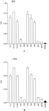
Figure 1
Figure 1
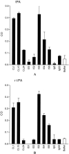
Identification of monoclonal anti-tPA Ab. A, some patient-derived monoclonal aCL bind to tPA. B, all monoclonal anti-tPA Ab bind to rtPA. Microtiter wells were coated with tPA or rtPA, and the test mAb, normal human IgG or the monoclonal isotype controls (IgG1 or IgG3) were analyzed at 1 μg/ml. Except for IS1 and IS2 (which are IgG1), all mAb are IgG3. Bound IgG were measured and expressed in OD, and the mean and standard error of the mean (SEM) are given (n = 2).
Figure 2
Figure 2
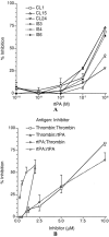
Competitive inhibition of mAb binding to tPA. The results are expressed in % inhibition, and the mean and SEM are given (n = 2). Crossreactivity of CL15 to rtPA and human α-thrombin (B). Binding of CL15 to either rtPA or thrombin in a solid phase was inhibited by soluble antigens (rtPA or thrombin). The experiment was performed in a similar manner to that in A except the cross-inhibition. The results are expressed in % inhibition, and the mean and SEM are given (n = 2-5).
Figure 3
Figure 3
Reactivity of monoclonal anti-tPA Ab with tPA and rtPA on fibrin surface (designated tPA-fibrin and rtPA-fibrin, respectively). All 6 anti-tPA mAb bind to tPA-fibrin (panel A) and 5/6 anti-tPA mAb bind to rtPA-fibrin (panel B). Notably, all 3 mAb with higher affinity to tPA (i.e., CL1, CL15 and IS3) bind stronger to tPA-fibrin than the other mAb.
Figure 4
Figure 4
Presence of IgG anti-tPA-fibrin Ab in some APS patients. A, plasma samples from 80 APS patients, 81 SLE patients, and 28 normal controls were analyzed at the 1:25 dilution. During each assay, CL15 at 1 μg/ml was used in each ELISA plate to serve as the reference sample, and the OD of each test sample was divided by that of CL15 in the same plate, resulting in a reference unit (RU) for each sample. B, the same data are normalized against the IgG level of each sample and are expressed in RU/IgG. The horizontal bars indicate the mean RU or mean RU/IgG for each group; the dashed line represents the cutoff, which is mean RU (or mean RU/IgG) plus 2 SD of the 28 normal controls. A representative result from three experiments is shown. A significant difference between the groups was denoted as * (p < 0.05) or ** (p < 0.01).
Figure 5
Figure 5
CL1 and CL15 inhibit tPA activity on fibrin surface. Test mAb and control IgG (at 100 μg/ml) were mixed with 100 nM rtPA for 15 minutes at room temperature, and then the mixtures were distributed to fibrin-coated wells. After incubation for one hour and wash, a tPA substrate S2765 was added and OD450 was measured after two hours. The data are expressed in OD change (A) and % inhibition (B) from normal human IgG control. CL15 inhibits tPA activity on fibrin surface in a concentration-dependent manner (C). The mean and SEM are given (n = 8). * denotes a significant difference from normal human IgG control (p < 0.05).

Similar articles

Cited by

References

    1. Harris EN, Pierangeli SS. Antiphospholipid antibodies and the antiphospholipid syndrome. Springer Semin Immunopathol. 1994;16:223–45. - PubMed
    1. Matsuura E, Igarashi Y, Fujimoto M, Ichikawa K. Anticardiolipin cofactor(s) and differential diagnosis of autoimmune disease. Lancet. 1990;336:177–8. - PubMed
    1. McNeil HP, Simpson RJ, Chesterman CN, Krilis SA. Anti-phospholipid antibodies are directed against a complex antigen that includes a lipid binding inhibitor of coagulation: beta 2-glycoprotein I (apolipoprotein H) Proc Natl Acad Sci USA. 1990;87:4120–4. - PMC - PubMed
    1. Galli M, Comfurius P, Maassen C, Hemker HC, de Baets MH, van Breda-Vriesman PJC, Barbui T, Zwaal RFA, Bevers EM. Anticardiolipin antibodies (ACA) directed not to cardiolipin but to a plasma protein cofactor. Lancet. 1990;335:1544–7. - PubMed
    1. Fleck RA, Rapaport SI, Rao LV. Anti-prothrombin antibodies and the lupus anticoagulant. Blood. 1988;72:512–9. - PubMed

Publication types

MeSH terms