Review
. 2005 Dec 29;360(1464):2201-10.
doi: 10.1098/rstb.2005.1760.

The oxygen sensing signal cascade under the influence of reactive oxygen species

Affiliations
Review

The oxygen sensing signal cascade under the influence of reactive oxygen species

Helmut Acker. Philos Trans R Soc Lond B Biol Sci. .

Abstract

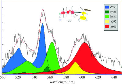
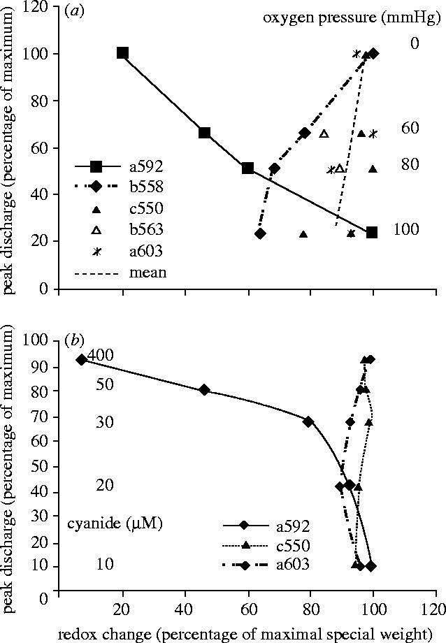
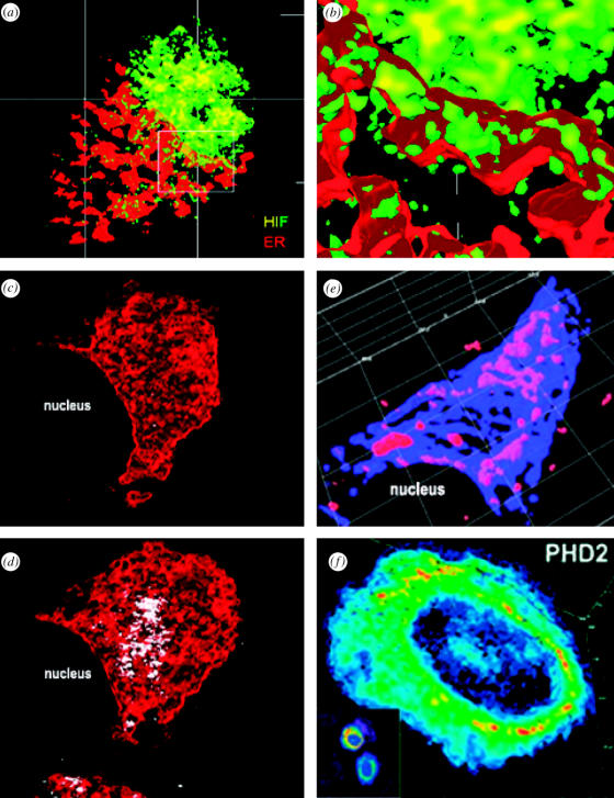
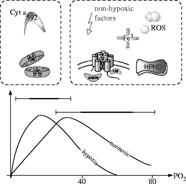
Structural and functional integrity of organ function profoundly depends on a regular oxygen and glucose supply. Any disturbance of this supply becomes life threatening and may result in severe loss of organ function. Particular reductions in oxygen availability (hypoxia) caused by respiratory or blood circulation irregularities cannot be tolerated for longer periods due to an insufficient energy supply by anaerobic glycolysis. Complex cellular oxygen sensing systems have evolved to tightly regulate oxygen homeostasis. In response to variations in oxygen partial pressure (PO2), these systems induce adaptive and protective mechanisms to avoid or at least minimize tissue damage. These various responses might be based on a range of oxygen sensing signal cascades including an isoform of the neutrophil NADPH oxidase, different electron carrier units of the mitochondrial chain such as a specialized mitochondrial, low PO2 affinity cytochrome c oxidase (aa3) and a subfamily of 2-oxoglutarate dependent dioxygenases termed HIF (hypoxia inducible factor) prolyl-hydroxylase and HIF asparaginyl hydroxylase called factor-inhibiting HIF (FIH-1). Thus, specific oxygen sensing cascades involving reactive oxygen species as second messengers may by means of their different oxygen sensitivities, cell-specific and subcellular localization help to tailor various adaptive responses according to differences in tissue oxygen availability.


Figures

Figure 1
Figure 1
Identification of carotid body heme proteins by light absorption photometry. N2 versus aerobic steady state spectrum (black solid noisy line) as a mean of six carotid bodies fitted by different mitochondrial and non-mitochondrial cytochrome spectra as indicated by different colours. The superposition curve (red solid line) obtained by varying the amplitude of the optical density of five cytochromes closely fits the experimental curve (Streller et al. 2002).
Figure 2
Figure 2
Nonlinear redox change of putative oxygen sensing heme proteins. (a) Redox changes of different carotid body heme proteins as calculated by deconvolution as shown in figure 1 are related to different PO2 values in the superfusion bath of the isolated haemoglobin free carotid body as well as to the chemoreceptor peak discharge. Cytochromes a592 and b558 only show a nonlinear redox change fitting the PO2–chemoreceptor discharge relationship. (b) Redox changes of different carotid body heme proteins as calculated by deconvolution as shown in figure 1 are related to different CN concentrations in the superfusion bath of the isolated haemoglobin free carotid body as well as to the chemoreceptor peak discharge. Cytochrome a592 only shows a nonlinear redox change. B type cytochromes are not detectable by deconvolution due to CN insensitivity (Streller et al. 2002).
Figure 3
Figure 3
Three-dimensional 2-photon confocal microscopy of components involved in hypoxia induced gene expression. (a,b) Translocation of HIF-1α (yellow) from the ER (red) into the nucleus of a human liver tumour cell (HepG2) under hypoxia (Berchner-Pfannschmidt et al. 2004). (c,d) OH. production (white) in the ER (red) of a human liver tumour cell (HepG2) under hypoxic (c) and normoxic (d) conditions. OH. production was stimulated by a 5 s blue light illumination inducing a photoreduction of FAD containing oxidases (Liu et al. 2004). (e) Overexpression of p22phox (blue) of the NADPH oxidase in the perinuclear space of a human liver tumour cell (HepG2) with an increased OH. production (red) probably fuelling an ER-based Fenton reaction (Djordjevic et al. 2005). (f) Predominant perinuclear localization of PHD2 in a human osteosarcoma cell (U2OS) (Metzen et al. 2003).
Figure 4
Figure 4
Modulation of oxygen sensor sensitivity by co-factors. Oxygen sensing systems connecting an oxygen-dependent enzymatic activity to the regulation of hypoxia-inducible responses should operate at high and low PO2 affinities, fitting the heterogeneous PO2 distribution curve (lower part). Oxygen sensing heme proteins such as mitochondrial complex IV (cytochrome c oxidase) and NAD(P)H-oxidase as well as PHD (upper part) have been described as candidate sensor systems functioning at different Km for PO2. Modulation of the specific PO2 affinities or oxygen sensor activities would allow to efficiently trigger and fine-tune various signal cascades to optimize cellular function and adaptation over a broad range of O2 concentrations (Acker & Acker 2004).

References

    1. Acker T, Acker H. Cellular oxygen sensing need in CNS function: physiological and pathological implications [Review] J. Exp. Biol. 2004;207:3171–3188. 10.1242/jeb.01075 - DOI - PubMed
    1. Acker H, Lübbers D.W. The kinetics of local tissue PO2 decrease after perfusion stop within the carotid body of the cat in vivo and in vitro. Pflüg. Arch. 1977;369:135–140. 10.1007/BF00591569 - DOI - PubMed
    1. Acker H, Lübbers D.W, Purves M.J, Tan E.D. Measurements of the partial pressure of oxygen in the carotid body of fetal sheep and newborn lambs. J. Dev. Physiol. 1980;2:323–328. - PubMed
    1. Archer S.L, Reeve H.L, Michelakis E, Puttagunta L, Waite R, Nelson D.P, Dinauer M.C, Weir E.K. O2 sensing is preserved in mice lacking the gp91 phox subunit of NADPH oxidase. Proc. Natl Acad. Sci. USA. 1999;96:7944–7949. 10.1073/pnas.96.14.7944 - DOI - PMC - PubMed
    1. Arthur P.G, Ngo C.T, Moretta P, Guppy M. Lack of oxygen sensing by mitochondria in platelets. Eur. J. Biochem. 1999;266:215–219. 10.1046/j.1432-1327.1999.00846.x - DOI - PubMed

MeSH terms

LinkOut - more resources