Review
. 2005 Dec 29;360(1464):2309-14.
doi: 10.1098/rstb.2005.1766.

The role of an astrocytic NADPH oxidase in the neurotoxicity of amyloid beta peptides

Affiliations
Review

The role of an astrocytic NADPH oxidase in the neurotoxicity of amyloid beta peptides

Andrey Y Abramov et al. Philos Trans R Soc Lond B Biol Sci. .

Abstract

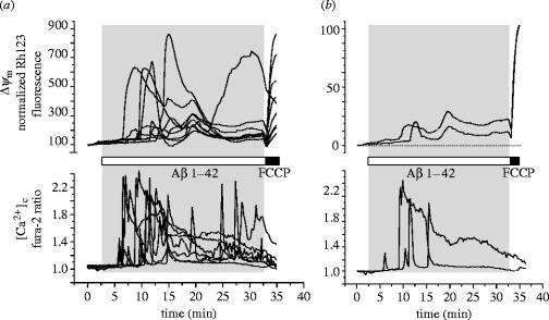
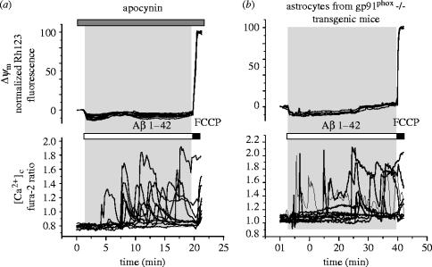
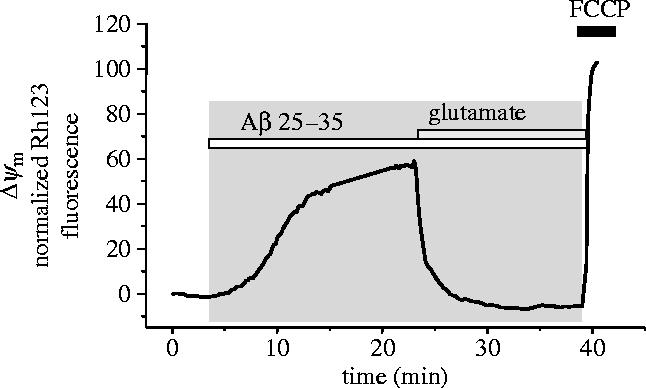
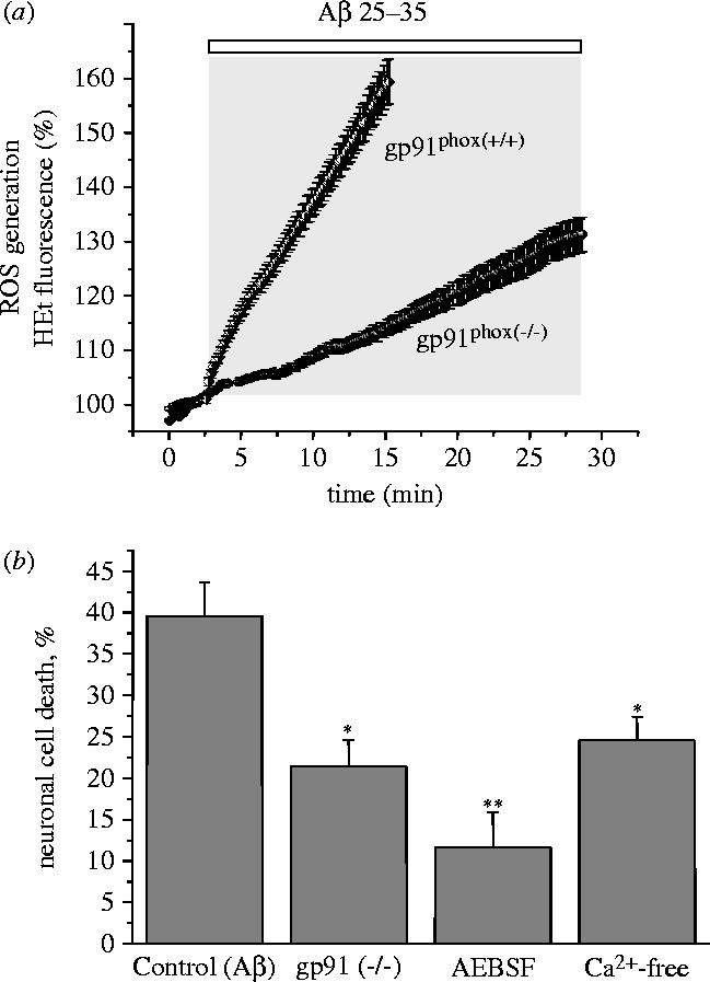
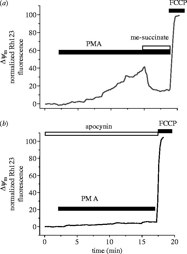
Amyloid beta peptide (Abeta) accumulates in the CNS in Alzheimer's disease. Both the full peptide (1-42) or the 25-35 fragment are toxic to neurons in culture. We have used fluorescence imaging technology to explore the mechanism of neurotoxicity in mixed asytrocyte/neuronal cultures prepared from rat or mouse cortex or hippocampus, and have found that Abeta acts preferentially on astrocytes but causes neuronal death. Abeta causes sporadic transient increases in [Ca2+]c in astrocytes, associated with a calcium dependent increased generation of reactive oxygen species (ROS) and glutathione depletion. This caused a slow dissipation of mitochondrial potential on which abrupt calcium dependent transient depolarizations were superimposed. The mitochondrial depolarization was reversed by mitochondrial substrates glutamate, pyruvate or methyl succinate, and by NADPH oxidase (NOX) inhibitors, suggesting that it reflects oxidative damage to metabolic pathways upstream of mitochondrial complex I. The Abeta induced increase in ROS and the mitochondrial depolarization were absent in cells cultured from transgenic mice lacking the NOX component, gp91phox. Neuronal death after 24 h of Abeta exposure was dramatically reduced both by NOX inhibitors and in gp91phox knockout mice. Thus, by raising [Ca2+]c in astrocytes, Abeta activates NOX, generating oxidative stress that is transmitted to neurons, causing neuronal death.


Figures

Figure 1
Figure 1
Aβ causes transient calcium signals, transient mitochondrial depolarizations, and a slow progressive loss of mitochondrial potential in mouse astrocytes. A culture of hippocampal cortical astrocytes was co-loaded with fura-2 (AM ester, 5 μM for 20 min) and with rhodamine 123 (100 μM for 15 min followed by washing). Application of Aβ 1–42 (2 μM) as indicated initiated transient calcium responses and also transient mitochondrial depolarizations which were superimposed on a slow progressive loss of mitochondrial potential. (a) Signals from many cells are superimposed. (b) Signals from just two cells are extracted so that individual cellular responses can be seen more clearly. These cellular responses in mouse astrocytes were not notably different from those we have described in cells from the rat (n=67 cells).
Figure 2
Figure 2
The changes in mitochondrial potential caused by Aβ were attributable to the activation of an NADPH oxidase. (a) The NADPH oxidase inhibitor apocynin (1 mM) prevented any significant change in mitochondrial potential in response to Aβ but had no effect at all on the Aβ induced calcium signals (n=84 cells) in mouse cortical astrocytes. Similar responses were seen to the other NADPH oxidase inhibitors AEBSF (20 μM, n=44 cells) and DPI (0.5 μM; n=111 cells). (b) Astrocytes cultured from the cortex of transgenic mice with knockout of gp91phox expression also showed no mitochondrial response to Aβ while the calcium response was unaffected (n=97 cells).
Figure 3
Figure 3
The slow loss of mitochondrial potential was reversed by mitochondrial substrate. In some cells, the slow progressive collapse of mitochondrial potential was the dominant response to Aβ. This loss of potential was reversed by provision of mitochondrial substrate, in the case illustrated, by glutamate (1 mM).
Figure 4
Figure 4
Aβ induced generation of oxygen free radicals and cell death are largely attributable to NADPH oxidase. (a) The rate of ROS generation was measured using hydroethidine (HEt), which is oxidized to a fluorescent product by ROS. The rate of appearance of the fluorescent product was clearly increased in mouse astrocytes exposed to Aβ 1–42 (2.5 μM; n=59) which caused a 3.6 fold increase in the rate of rise of the HEt signal (from 1.1±0.12 to 3.98±0.21 arb. fluorescence U min−1; n=179 cells), in cortical astrocytes of both rats and mice. In astrocytes cultured from gp91phox knockout transgenic mice, no significant increase in ROS generation was detectable (n=116). (b) Neuronal death of cells from gp91phox knockout mice following 24 h exposure to Aβ was significantly reduced from control (39.6±4.1%) to 21.4±3.2% (*p<0.05). The inhibitor of NADPH oxidase AEBSF and removal of external Ca2+ both also reduced neuronal cell death in mouse cultures, (cell death was reduced from 39.6±4.1% in control to 11.6±4.3% (AEBSF, **p<0.001) and to 24.6±2.8% (Ca2+-free, *p<0.05).
Figure 5
Figure 5
Activation of the NADPH oxidase by PMA causes a slow loss of mitochondrial potential that is reversed by mitochondrial substrate. The ‘classical’ activator of the NADPH oxidase in neutrophils, PMA, increases ROS generation in astrocytes (not shown) and produced a slow but sustained mitochondrial depolarization in the majority of cortical astrocytes tested (n=91 of 98 cells). B. This effect was inhibited by apocynin (n=65) or DPI (0.5 μM; n=48, not shown). Further similarity between the Aβ- and PMA-induced changes in Δψm can be seen by the reversal of the PMA induced response by delivery of mitochondrial substrate—illustrated here for methyl-succinate. Similar data were obtained for glutamate (1 mM; n=62 for PMA and n=218 for Aβ) and methyl-succinate (n=66 for PMA and n=145 for Aβ).

References

    1. Abramov A.Y, Canevari L, Duchen M.R. Changes in intracellular calcium and glutathione in astrocytes as the primary mechanism of amyloid neurotoxicity. J. Neurosci. 2003;23:5088–5095. - PMC - PubMed
    1. Abramov A.Y, Canevari L, Duchen M.R. Amyloid β peptides induce mitochondrial dysfunction and oxidative stress in astrocytes and death of neurons through activation of NADPH oxidase. J. Neurosci. 2004a;23:5088–5095. - PMC - PubMed
    1. Abramov A.Y, Canevari L, Duchen M.R. Calcium signals induced by amyloid beta peptide and their consequences in neurons and astrocytes in culture. Biochim. Biophys. Acta. 2004b;6:81–87. - PubMed
    1. Abramov A.Y, Jacobson J, Wientjes F, Hothersall J, Canevari L, Duchen M.R. Expression and modulation of an NADPH oxidase in mammalian astrocytes. J. Neurosci. 2005;25:9176–9184. - PMC - PubMed
    1. Arispe N, Doh M. Plasma membrane cholesterol controls the cytotoxicity of Alzheimer's disease AβP (1–40) and (1–42) peptides. FASEB J. 2002;16:1526–1536. 10.1096/fj.02-0829com - DOI - PubMed

Publication types

LinkOut - more resources