Review
. 2005 Dec 29;360(1464):2355-72.
doi: 10.1098/rstb.2005.1770.

Oxidative stress and ageing: is ageing a cysteine deficiency syndrome?

Affiliations
Review

Oxidative stress and ageing: is ageing a cysteine deficiency syndrome?

Wulf Dröge. Philos Trans R Soc Lond B Biol Sci. .

Abstract

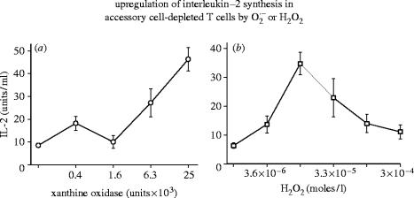
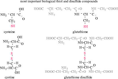
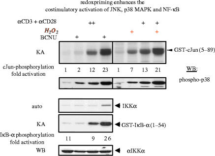
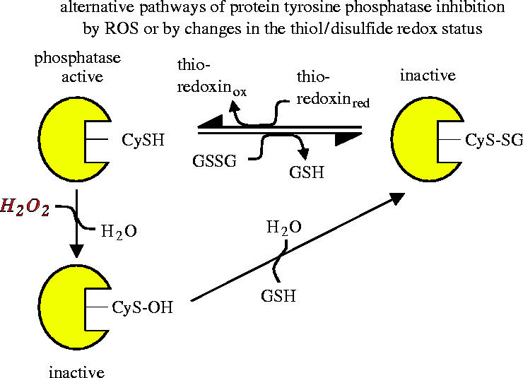
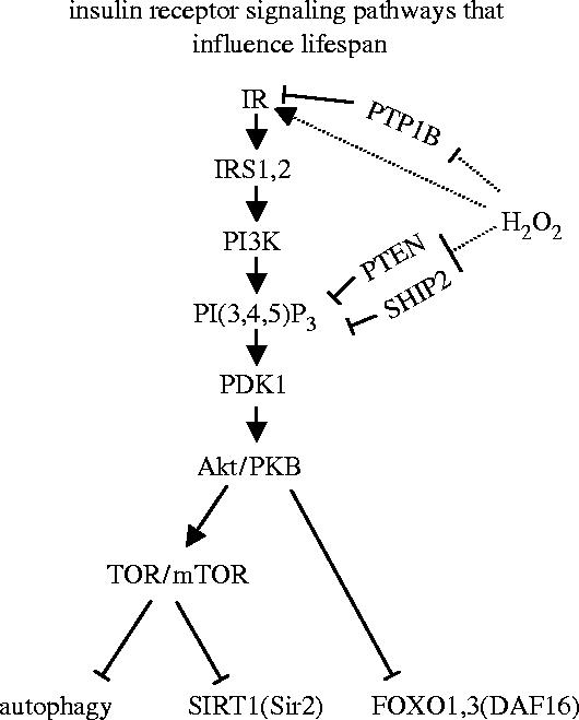
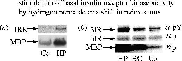
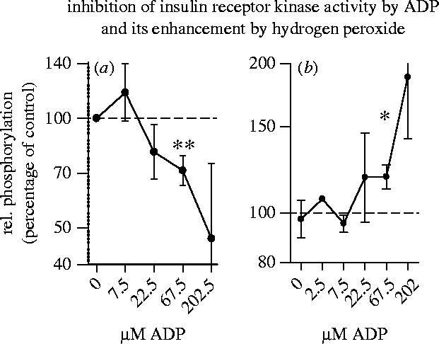
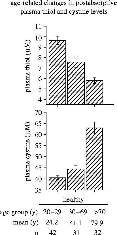
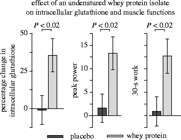
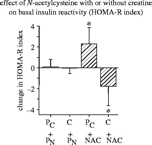
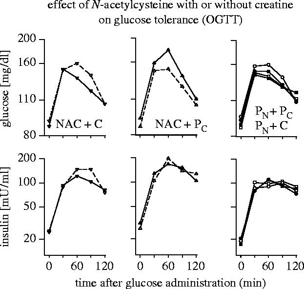
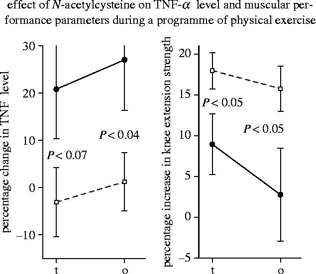
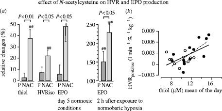
Reactive oxygen species (ROS) are constantly produced in biological tissues and play a role in various signalling pathways. Abnormally high ROS concentrations cause oxidative stress associated with tissue damage and dysregulation of physiological signals. There is growing evidence that oxidative stress increases with age. It has also been shown that the life span of worms, flies and mice can be significantly increased by mutations which impede the insulin receptor signalling cascade. Molecular studies revealed that the insulin-independent basal activity of the insulin receptor is increased by ROS and downregulated by certain antioxidants. Complementary clinical studies confirmed that supplementation of the glutathione precursor cysteine decreases insulin responsiveness in the fasted state. In several clinical trials, cysteine supplementation improved skeletal muscle functions, decreased the body fat/lean body mass ratio, decreased plasma levels of the inflammatory cytokine tumour necrosis factor alpha (TNF-alpha), improved immune functions, and increased plasma albumin levels. As all these parameters degenerate with age, these findings suggest: (i) that loss of youth, health and quality of life may be partly explained by a deficit in cysteine and (ii) that the dietary consumption of cysteine is generally suboptimal and everybody is likely to have a cysteine deficiency sooner or later.


Figures

Figure 1
Figure 1
Upregulation of interleukin-2 (IL-2)synthesis in accessory cell-depleted T cells by O2· or H2O2. IL-2 production in Concanavalin-A-stimulated accessory cell-depleted T cell populations in the presence of an O2-generating system (left panel) or hydrogen peroxide (right panel). For experimental details see Roth & Dröge (1987).
Figure 2
Figure 2
Redox-responsive transcription factors (schemtatic illustration). Nuclear factor-κB (NF-κB) is a p65/p50 heterodimer. It was the first transcription factor shown to be activated by ROS (see text). The activator protein-1 (AP-1) typically consists of Fos and Jun proteins and is activated under oxidative conditions through several redox-responsive signalling proteins upstream of N-termal Jun kinase (JNK).
Figure 3
Figure 3
Most important biological thiol and disulfide compounds of low molecular weight. Cysteine is a thiol-containing amino acid, which can be oxidized to the corresponding disulfide compound cystine. Glutathione is a tripeptide, consisting of glutamate, cysteine and glycine. Glutathione disulfide is formed by oxidation of glutathione.
Figure 4
Figure 4
Effects of BCNU on redox status and AP-1 activity. (a) Human T lineage cells (Molt-4) were treated with the glutathione reductase inhibitor BCNU at the indicated concentrations. After 3 h the cells were harvested, intracellular concentrations of total glutathione and glutathione disulfide were determined, and reduced glutathione (GSH) was calculated from the difference. (b) Molt-4 cells were transiently transfected with a CAT reporter construct driven by the thymidine kinase promoter under the control of 6 AP-1 binding sites. One day later the cells were treated with BCNU at the indicated concentrations and with or without TPA (10 ng ml−1). Transactivation was determined after 36 h by CAT assay. (For other details see Galter et al. 1994.)
Figure 5
Figure 5
Redox priming enhances the costimulatory activation of JNK, p38 MAPK and NF-κB. Jurkat cells were incubated with or without BCNU (10 μM) or hydrogen peroxide (30 μM) and activated with or without agonistic anti-CD3/anti-CD28 antibodies. Upper panels: Cells were lysed and tested for cJun phosphorylation by immune complex kinase assays and for p38 MAPK phosphorylation by immunoblotting. Lower panels: Endogenous IκB kinase α (IKKα) was immunoprecipitated and analysed either for its kinase activity (KA), using recombinant GST-IκB-α as a substrate or by Western blotting (WB) for the occurrence of IKKα. A longer exposure of the gel displaying the phosphorylation of IKK is also shown (Auto). (For other details see Hehner et al. 2000.)
Figure 6
Figure 6
Alternative pathways of protein tyrosine phosphatase inhibition by ROS or by changes in the thiol/disulfide redox status. A cysteine residue in the catalytic site of the phosphatase is critical for its catalytic activity. Inactivation can occur either by reaction with hydrogen peroxide to form a sulfenic acid derivative or by reaction with glutathione disulfide, resulting in glutathionylation of the critical cysteine residue. Reactivation of the phosphatase may occur by reaction with reduced glutathione or other thiol compounds (schematic illustration based on Barford et al. 1995; Barrett et al. 1999a,b).
Figure 7
Figure 7
Insulin receptor signalling pathways that influence life span. Binding of insulin leads to autophosphorylation and activation of the insulin receptor kinase (IR) and recruitment of insulin-receptor substrates (IRS-1, IRS-2). Phosphatidylinositol 3-kinase (PI3K) converts phosphatidylinositol(4,5)diphosphate (PI(4,5)P2) into phosphatidylinositol(3,4,5)triphosphate (PI(3,4,5)P3). This binds and activates phosphoinositide-dependent protein kinase 1 (PDK1), which phosphorylates the serine/threonine kinase Akt1. Akt1 is activated by binding to PI(3,4,5)P3 at the cell membrane and by phosphorylation. Akt1 stimulates the target of rapamycin (TOR or the mammalian mTOR) and protein synthesis but is an inhibitor of several other functions, including autophagy, SIRT1 activity and FOXO transcription factor activity. Insulin receptor signalling activity is downregulated by protein tyrosine phosphatase 1B (PTB1B), which dephosphorylates the IRK domain, and by the phosphatase and tensine homologue on chromosome 10 (PTEN) and SH2-domain-containing inositol phosphatase (SHIP2) both of which dephosphorylate PI(3,4,5)P3. Hydrogen peroxide enhances the autophosphorylation of IRK and inhibits the activity of the three phosphatases.
Figure 8
Figure 8
Stimulation of basal insulin receptor kinase activity by hydrogen peroxide or a shift in glutathione redox status. (a) Phosphorylation of recombinant IRK protein and myelin basic protein (MBP) substrate. The recombinant IRK protein was incubated at 30 °C for 20 min with or without 60 μM hydrogen peroxide (HP), then for 30 min with MBP and 1 mM ATP, and finally for 20 min with 32P-ATP and MBP. (b) Intact CHO-HIR cells were cultured with 50 μM hydrogen peroxide (HP), 80 μM 1-(2-chloroethyl)-3-(2-hydroxyethyl)-1-nitrosourea, a specific inhibitor of glutathione reductase (BC), or no additives (Co). The insulin receptor was purified by immunoprecipitation and assayed for autophosphorylation (βIR) or substrate phosphorylation (MBP) by 32P-incorporation or phosphotyrosine antibody (α-PY; data taken from Schmitt et al. 2005).
Figure 9
Figure 9
Inhibition of insulin receptor kinase activity by ADP and its enhancement by hydrogen peroxide. Recombinant IRK protein was incubated with or without 60 μM hydrogen peroxide and with or without the indicated concentrations of ADP. (a) shows the relative phosphorylation of myelin basic protein (MBP) substrate in cultures without hydrogen peroxide. (b) shows the relative enhancement of MBP phosphorylation by hydrogen peroxide as compared to controls without hydrogen peroxide. (For other details see Schmitt et al. 2005.)
Figure 10
Figure 10
Age-related changes in postabsorptive plasma thiol and cystine levels. Postabsorptive plasma amino acid and acid-soluble thiol levels have been determined in the plasma from the cubital vein of randomly selected healthy human subjects of both sexes (unpublished data from Wulf Hildebrandt, see also Hack et al. 1998).
Figure 11
Figure 11
Effect of undenatured whey protein on intracellular glutathione and muscle functions. The data indicate the relative change (%) in the intracellular total glutathione concentration of lymphocytes, peak power, and 30 s work capacity of healthy young subjects during a three month treatment and observation period. (For other details see Lands et al. 1999.)
Figure 12
Figure 12
Effect of N-acetylcysteine (NAC) with or without creatine (C) on basal insulin reactivity. The homeostasis model assessment/insulin resistance index (HOMA-R) was used as a widely accepted measure of basal insulin reactivity (Matthews et al. 1985). It was calculated by the formula: HOMA-R=fasting plasma glucose (mg dl−1)×fasting plasma insulin concentration (mU ml−1)×405−1. The data show the absolute changes (mean±s.e.m.) in the HOMA-R index between baseline and terminal examination in the four treatment groups: Pc/Pn=placebocreatine+placeboNAC,C/Pn=creatine+placeboNAC,Pc/NAC=placebocreatine+NACandC/NAC=creatine+NAC. (For other details see Hildebrandt et al. 2004.)
Figure 13
Figure 13
Effect of N-acetylcysteine with or without creatine on glucose tolerance (OGTT). Non-diabetic obese subjects have been treated with a combination of N-acetylcysteine+creatine (NAC+C; left panel), N-acetylcysteine+placebocreatine (middle panel), placeboNAC+creatine, or with placeboNAC+placebocreatine (right panel). The data show glucose and insulin concentrations at different times after glucose administration before (open symbols) and after the intervention and observation period (closed symbols). (For other details see Hildebrandt et al. 2004.)
Figure 14
Figure 14
Effect of N-acetylcysteine on TNF-α level and muscular performance during a programme of physical exercise. The data indicate the relative increase (%) in plasma TNF-α level and knee extension strength during a six week treatment and exercise programme (t), and during the total 12–13 week observation period (o). Filled circles, placebo group; open squares, N-acetylcysteine-treated group (see Hauer et al. 2003).
Figure 15
Figure 15
Effect of N-acetylcysteine on hypoxic ventilatory response (HVR) and erythropoietin (Epo) production. (a) Relative changes during medication (N-acetylcysteine, NAC) between baseline and terminal examination expressed as percentage of baseline values. The data show the changes in plasma thiol concentrations, HVR under isocapnic conditions, EPO concentration under nomoxic conditions, and EPO 2 h after exposure to prolonged normobaric hypoxia. (b) Shows the correlation between the poikilocapnic HVR with the corresponding plasma thiol level. Each point represents one subject (filled circle, N-acetylcysteine-treated; open circle, placebo group). The solid line shows the regression function of the total population (r=0.59, P<0.01). (For other details see Hildebrandt et al. 2002a,.)

Similar articles

Cited by

References

    1. Acharyya S, Ladner K.J, Nelsen L.L, Damrauer J, Reiser P.J, Swoap S, Guttridge D.C. Cancer cachexia is regulated by selective targeting of skeletal muscle gene products. J. Clin. Invest. 2004;114:370–378. 10.1172/JCI200420174 - DOI - PMC - PubMed
    1. Arivazhagan P, Ramanathan K, Panneerselvam C. Effect of dl-alpha-lipoic acid on mitochondrial enzymes in aged rats. Chem. Biol. Interact. 2001;138:189–198. 10.1016/S0009-2797(01)00268-X - DOI - PubMed
    1. Azzoni L, Papasavvas E, Chehimi J, Kostman J.R, Mounzer K, Ondercin J, Perussia B, Montaner L.J. Sustained impairment of IFN-γ secretion in suppressed HIV-infected patients despite mature NK cell recovery: evidence for a defective reconstitution of innate immunity. J. Immunol. 2002;168:5764–5770. - PubMed
    1. Balkan J, Kanbagli O, Mehmetcik G, Mutlu-Turkoglu U, Aykac-Toker G, Uysal M. Increased lipid peroxidation in serum and low-density lipoproteins associated with aging in humans. Int. J. Vitam. Nutr. Res. 2002;72:315–320. - PubMed
    1. Barford D, Jia Z, Tonks N.K. Protein tyrosine phosphatases take off. Nat. Struct. Biol. 1995;2:1043–1053. 10.1038/nsb1295-1043 - DOI - PubMed

MeSH terms