. 2005 Dec;187(24):8411-26.
doi: 10.1128/JB.187.24.8411-8426.2005.

The ClgR protein regulates transcription of the clpP operon in Bifidobacterium breve UCC 2003

Affiliations

The ClgR protein regulates transcription of the clpP operon in Bifidobacterium breve UCC 2003

Marco Ventura et al. J Bacteriol. 2005 Dec.

Abstract

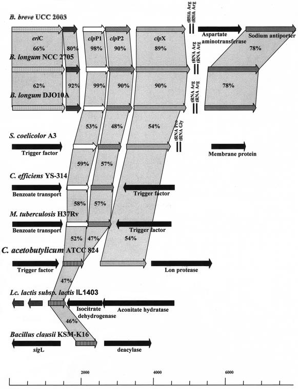
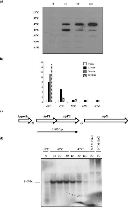
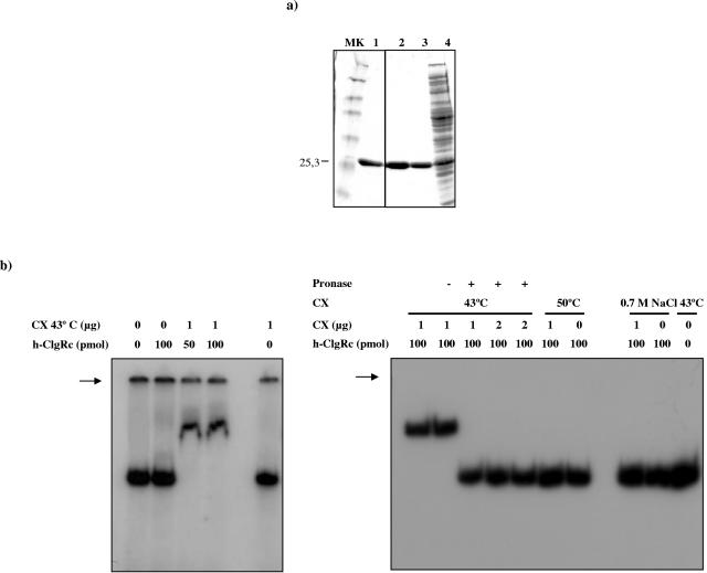
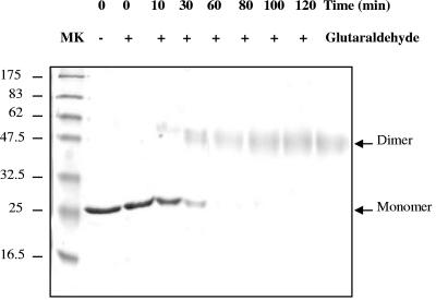
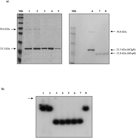
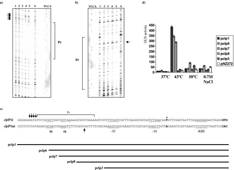
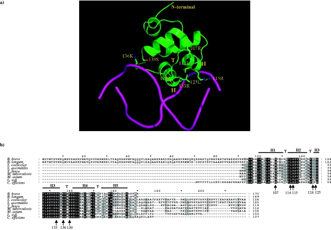
Five clp genes (clpC, clpB, clpP1, clpP2, and clpX), representing chaperone- and protease-encoding genes, were previously identified in Bifidobacterium breve UCC 2003. In the present study, we characterize the B. breve UCC 2003 clpP locus, which consists of two paralogous genes, designated clpP1 and clpP2, whose deduced protein products display significant similarity to characterized ClpP peptidases. Transcriptional analyses showed that the clpP1 and clpP2 genes are transcribed in response to moderate heat shock as a bicistronic unit with a single promoter. The role of a clgR homologue, known to control the regulation of clpP gene expression in Streptomyces lividans and Corynebacterium glutamicum, was investigated by gel mobility shift assays and DNase I footprint experiments. We show that ClgR, which in its purified form appears to exist as a dimer, requires a proteinaceous cofactor to assist in specific binding to a 30-bp region of the clpP promoter region. In pull-down experiments, a 56-kDa protein copurified with ClgR, providing evidence that the two proteins also interact in vivo and that the copurified protein represents the cofactor required for ClgR activity. The prediction of the ClgR three-dimensional structure provides further insights into the binding mode of this protein to the clpP1 promoter region and highlights the key amino acid residues believed to be involved in the protein-DNA interaction.


Figures

FIG. 1.
FIG. 1.
Comparison of the clpP operon in B. breve UCC 2003 and corresponding loci in various other bacteria. Each arrow indicates an open reading frame. The lengths of the arrows are proportional to the lengths of the predicted open reading frames. Orthologs are marked with the same color. The putative function of the protein is indicated above each arrow. The amino acid identity of each protein with respect to B. breve UCC 2003 is indicated as a percentage.
FIG. 2.
FIG. 2.
Phylogenetic tree obtained using the clpP1 and clpP2 gene sequences. Bar, phylogenetic distances. Bootstrap values are reported for a total of 1,000 replicates. The clpP1 and clpP2 gene sequences are indicated. Bacteria belonging to the Firmicutes and Actinobacteridae groups are indicated.
FIG. 3.
FIG. 3.
Transcriptional analysis of the B. breve UCC 2003 clpP operon. (a) Slot blot hybridization using RNAs extracted from cells incubated for up to 150 min at various temperatures or with various NaCl concentrations (indicated in the left-hand margin). (b) Schematic representation of mRNA levels of induction. The different colors and filling of the pillars correspond to the various times for which heat and osmotic shocks were applied, as indicated in the figure. (c) Position of transcript with respect to the clpP locus map. The estimated size of the transcript is indicated. Hairpin symbols indicate possible rho-independent terminators. (d) Northern blot analysis of the B. breve UCC 2003 clpP operon performed using clpP1 as a probe and total mRNA isolated from cultures exposed to 37°C or under heat or hyperosmotic conditions for the times indicated.
FIG. 4.
FIG. 4.
Organization of the clgR locus in different members of the Actinobacteridae. Each arrow indicates an open reading frame. The lengths of the arrows are proportional to the lengths of the predicted open reading frames. Orthologs are marked with the same color. The putative function of the protein is indicated above each arrow. The amino acid identity of each protein with respect to B. breve UCC 2003 is indicated as a percentage.
FIG. 5.
FIG. 5.
Detection of ClgR binding to the clpP1 promoter of B. breve UCC 2003. (a) Overproduction and purification of ClgR. SDS-PAGE analysis was performed with the purified ClgR protein (lanes 1 to 3, containing 15, 20, and 8 μg of protein, respectively) and with crude extracts from E. coli M15 carrying pEQ-ClgR upon IPTG induction (lane 4). A molecular weight standard (Bio-Rad, United Kingdom) was loaded in lane MK. (b) Gel retardation assays were performed with the clpP1p fragment as a probe. CX indicates a crude lysate from B. breve UCC 2003 cultures grown at 43°C or 50°C or in medium with 0.7 M NaCl. The amounts of h-ClgR and CX used are indicated above each lane. Gel retardation results for the clpP1p fragment as a probe and h-ClgR plus CX grown at 43°C, which prior to the binding experiment had been treated with pronase (+) or incubated in its absence, are presented in the rightmost panel. The arrows indicate the positions of the wells.
FIG. 6.
FIG. 6.
Subunit composition of ClgR. An SDS-PAGE gel of glutaraldehyde-cross-linked and non-cross-linked h-ClgR is shown. The absence (−) or presence (+) of the glutaraldehyde cross-linking reagent and the time of the cross-linking reaction are shown at the top of each lane. The positions of the h-ClgR monomer and dimer forms are indicated.
FIG. 7.
FIG. 7.
Pull-down assay to identify the cofactor of ClgR (a) and DNA binding assay using activated and nonactivated h-ClgR molecules (b). (a) Lanes 1 and 2, SDS-PAGE of h-ClgR coeluted with CX obtained from a culture grown at 43°C; lane 3, SDS-PAGE of h-ClgR coeluted with CX obtained from a culture grown at 50°C; lane 4, SDS-PAGE of h-ClgR eluted with CX obtained from cells grown in the presence of 0.7 M NaCl; lane 5, SDS-PAGE of purified h-ClgR (positive control); lane 6, SDS-PAGE of h-HspR coeluted with CX grown at 43°C; lane 7, SDS-PAGE of purified h-HspR (positive control). (b) DNA binding assays using h-ClgR molecules derived from copurification experiments. Lane 1, 100 pmol of h-ClgR derived from coelution with CX grown at 43°C; lane 2, 100 pmol of h-ClgR derived from coelution with CX grown at 43°C with 1 mM ATP; lane 3, 100 pmol of h-ClgR derived from coelution with CX grown at 50°C; lane 4, 100 pmol of h-ClgR derived from coelution with CX grown at 50°C with 1 mM ATP; lane 5, 100 pmol of h-ClgR derived from coelution with CX grown in medium containing NaCl at a concentration of 0.7 M; lane 6, 100 pmol of h-ClgR derived from coelution with CX grown at 50°C with 1 mM ATP; lane 7, no protein was used; lane 8, 100 pmol of h-ClgR plus 1 μg CX grown at 43°C (positive control).
FIG. 8.
FIG. 8.
DNase I footprints of the 347-bp fragment from the B. breve UCC 2003 clpP1 promoter region for the template strand (a) and the nontemplate strand (b). (c and d) β-Glucuronidase activities of various clpP1-gusA fusions in B. breve UCC 2003 grown at a range of temperatures or with 0.7 M NaCl. (a and b) Lane 1, no protein extract; lane 2, 100 pmol of h-ClgR; lane 3, 25 pmol of h-ClgR plus 1 μg of crude lysate from B. breve UCC 2003 cultures (CX) grown at 43°C; lane 4, 50 pmol of h-ClgR and 1 μg CX grown at 43°C; lane 5, 100 pmol of h-ClgR and 1 μg CX grown at 43°C; lane 6, 1 μg CX grown at 43°C. Pr, ClgR-protected region. Arrows indicate hypersensitivity sites. (c) Template (clpP1t) and nontemplate (clpP1nt) clpP1 promoter sequences. IR, putative regulator sequences. The −10 and −35 hexamers are underlined, letters show in bold denote the transcription start site, and arrows indicate hypersensitivity sites. Bars indicate the positions of the genetic constructs with respect to the clpP1 promoter region. (d) GUS activities of the pclp1, pclp6, pclp7, pclp8, and pclp3 transcription fusions and the empty vector pNZ272 grown at 37°C, 43°C, or 50°C or in medium containing 0.7 M NaCl.
FIG. 9.
FIG. 9.
Predicted 3D model for the C-terminal end of ClgR. (a) In order to predict the mode of binding between ClgR and the DNA molecule, the DNA molecule (the ligand) from the crystal structure of 1rio was superimposed onto the predicted ClgR model and is depicted by magenta tubes. Potential residues in ClgR involved in the interaction with the DNA molecule are indicated. The atoms in these highlighted residues are colored and are represented as sticks. Color code: green, carbon; red, oxygen; blue, nitrogen. (b) Alignment of ClgR proteins from several high-G+C gram-positive bacteria. Shading indicates conservation at a given position in at least 50% of the proteins in the alignment as either identical (black) or similar (light gray) residues. The amino acid residues involved in DNA binding are indicated with arrows, the turns are indicated with “T,” and the helices are indicated with “H.”

Similar articles

Cited by

References

    1. Ashok, K. D., C. S. Baker, K. Suzuki, A. D. Jones, P. Pandit, T. Romero, and P. Babitzke. 2003. CsrA regulates translation of the Escherichia coli carbon starvation gene cstA by blocking ribosome access to the cstA transcript. J. Bacteriol. 15:4450-4460. - PMC - PubMed
    1. Bellier, A., and P. Mazodier. 2004. ClgR, a novel regulator of clp and lon expression in Streptomyces. J. Bacteriol. 186:3238-3248. - PMC - PubMed
    1. Bucca, G., A. M. E. Brassington, H. J. Schonfeld, and C. P. Smith. 2000. The HspR regulon of Streptomyces coelicolor: a role for the DnaK chaperone as a transcriptional co-repressor. Mol. Microbiol. 38:1093-1103. - PubMed
    1. Crecy-Lagard, V., P. Servant-Moisson, J. Viala, C. Grandvalet, and P. Mazodier. 1999. Alteration of the synthesis of the Clp ATP-dependent protease affects morphological and physiological differentiation in Streptomyces. Mol. Microbiol. 32:505-517. - PubMed
    1. Derre, I., G. Rapoport, and T. Msadek. 1999. CtsR, a novel regulator of stress and heat shock response, controls clp and molecular chaperone gene expression in gram-positive bacteria. Mol. Microbiol. 31:117-131. - PubMed

Publication types

MeSH terms

Associated data

LinkOut - more resources