. 2005 Nov 30;33(21):6782-94.
doi: 10.1093/nar/gki979. Print 2005.

A genomic approach to the identification and characterization of HOXA13 functional binding elements

Affiliations

A genomic approach to the identification and characterization of HOXA13 functional binding elements

Colleen D McCabe et al. Nucleic Acids Res. .

Abstract

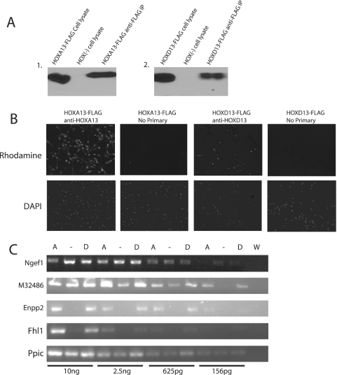
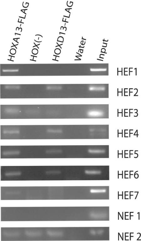
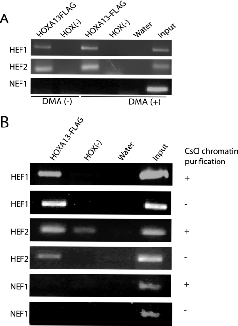
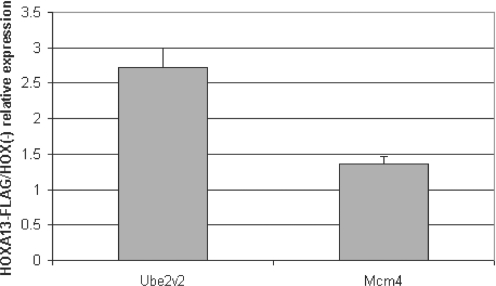
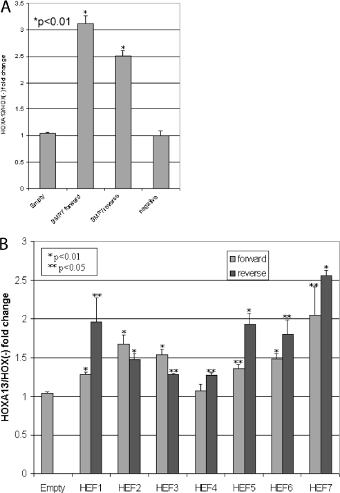
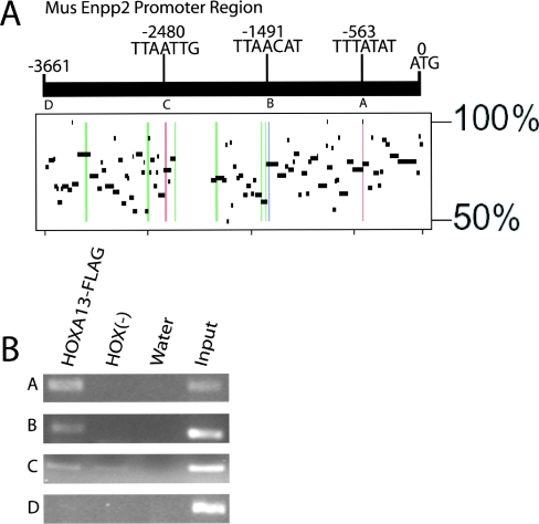
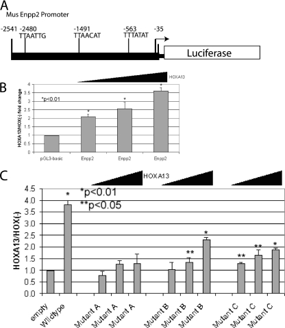
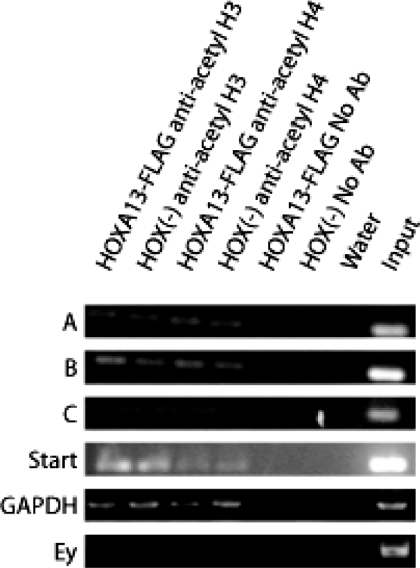
HOX proteins are important transcriptional regulators in mammalian embryonic development and are dysregulated in human cancers. However, there are few known direct HOX target genes and their mechanisms of regulation are incompletely understood. To isolate and characterize gene segments through which HOX proteins regulate transcription we used cesium chloride centrifugation-based chromatin purification and immunoprecipitation (ChIP). From NIH 3T3-derived HOXA13-FLAG expressing cells, 33% of randomly selected, ChIP clones were reproducibly enriched. Hox-enriched fragments (HEFs) were more AT-rich compared with cloned fragments that failed reproducible ChIP. All HEFs augmented transcription of a heterologous promoter upon coexpression with HOXA13. One HEF was from intron 2 of Enpp2, a gene highly upregulated in these cells and has been implicated in cell motility. Using Enpp2 as a candidate direct target, we identified three additional HEFs upstream of the transcription start site. HOXA13 upregulated transcription from an Enpp2 promoter construct containing these sites, and each site was necessary for full HOXA13-induced expression. Lastly, given that HOX proteins have been demonstrated to interact with histone deacetylases and/or CBP, we explored whether histone acetylation changed at Enpp2 upon HOXA13-induced activation. No change in the general histone acetylation state was observed. Our results support models in which occupation of multiple HOX binding sites is associated with highly activated genes.


Figures

Figure 1
Figure 1
Characterization of HOXA13-FLAG/EGFP or HOXD13-FLAG/EGFP expressing cells. (A) Western blot using HOX-specific antibodies demonstrating HOXA13 expression and anti-FLAG immunoprecipitation from HOXA13-FLAG cell line and absent HOXA13 expression in the HOX (−) cell line (1); HOXD13-FLAG expression and anti-FLAG immunoprecipitation from HOXD13-FLAG cell line but absent HOXD13 expression in the HOX (−) cell line (2). (B) Immunocytochemistry using Hox specific antibodies and DAPI staining demonstrate expression and nuclear localization of HOXA13 or HOXD13 in their respective cell lines. (C) Input RNA using serial dilutions ranging from 156 pg to 10 ng was used in semi-quantitative RT–PCR to look for expression changes of four reported targets. Fhl1 (+6-fold), Enpp2 (+18.8-fold) and M32486 (+2.3) are upregulated in the HOXA13-FLAG (A) and HOXD13-FLAG (D) cell lines and Ngef (−2.4) is downregulated compared to HOX (−). Water (W) was used as a PCR control. Ppic was a loading control and was shown to be unchanged in the HOXA13-FLAG and HOXD13-FLAG cell lines.
Figure 2
Figure 2
Representative PCR enrichment of HEFs. ChIP was performed in HOXA13-FLAG, HOX (−) and HOXD13-FLAG cell lines using anti-FLAG agarose. PCR detection was performed using primers specific to each HEF, NEF1 and NEF2. NEF1 resulted in no detectable product for each cellular sample and NEF2 resulted in product with no detectable difference between the HOXA13-FLAG or HOXD13-FLAG cell lines and the HOX (−) cells. Water was used as a negative PCR control and input ChIP DNA from the HOX (−) cells was used as the positive PCR control.
Figure 3
Figure 3
HEF enrichment without protein–protein crosslinking and chromatin purification. (A) ChIP was performed in HOXA13-FLAG and HOX (−) cells using anti-FLAG agarose. Elimination of crosslinking with DMA preceding formaldehyde crosslinking is represented in indicated lanes [DMA(−)]. HEF1 and HEF2 are enriched upon addition of DMA as well as without DMA in the HOXA13-FLAG versus HOX (−) cells. HEF1 demonstrated a visibly higher signal with DMA versus DMA (−) in the HOXA13-FLAG cells (1.9-fold by BioRad Quantity One analysis) while HEF2 recovery was equal between the samples. (B) ChIP was performed in HOXA13-FLAG and HOX (−) cell lines using anti-FLAG agarose. CsCl purification of chromatin was eliminated in the indicated samples (−). HEF1 and HEF2 are both enriched in the HOXA13-FLAG cells versus the HOX (−) cells both with and without chromatin purification. There was consistently no product in the HOX (−) sample for HEF1; however, there is a product present in the CsCl purified HEF2 sample.
Figure 4
Figure 4
Ube2v2 is upregulated 2.5-fold in HOXA13-FLAG cells. Input RNA isolated from HOXA13-FLAG and HOX (−) cell lines were used in real time PCR assays for Ube2v2 and Mcm4. Hprt was used as a loading control to normalize the values between cell lines. Ube2v2 expression was 2.5-fold upregulated in the HOXA13-FLAG cell versus the HOX (−) cells. Mcm4 expression was not changed between cell lines.
Figure 5
Figure 5
HOXA13 enhances transcription from HEFs. (A) The enhancer region of Bmp7 (20) was cloned upstream of the chicken β-actin minimal promoter driving luciferase and cotransfected in COS7 cells with CMV-HOXA13 or pCMV as a positive control for HOXA13 function. Addition of CMV-HOXA13 resulted in a 2.5- to 3.1-fold increase in activity over empty vector. NEF1 was also cloned upstream of the chicken β-actin minimal promoter driving luciferase and used in the same assay resulted in no detectable difference in normalized reporter activity upon addition of CMV-HOXA13 over empty vector. A β-galactosidase expression vector was cotransfected as a transfection control and all samples were normalized to its activity. (B) HEFs were cloned in both a forward and reverse orientation upstream of the chicken β-actin minimal promoter driving luciferase and cotransfected in COS7 cells with CMV-HOXA13 or pCMV. Luciferase reporter activity upon addition of HOXA13 resulted in a significant (*P < 0.01; **P < 0.05) increase in relative luciferase activity when compared with identical transfections with control pCMV vector. A β-galactosidase expression vector was used as a transfection control and all samples were normalized to lacZ activity.
Figure 6
Figure 6
Candidate HOXA13 binding sites in the Enpp2 upstream region are enriched in HOXA13-FLAG expressing cells. (A) Candidate in vivo binding sites for HOXA13 were identified upstream of the mouse Enpp2 translational start methionine (ATG) using in vitro core sequence variations (9,20) and are labeled with their position relative to the ATG. The plot resulting from an analysis using Advanced Pipmaker () shows sequence conservation to the region upstream of the human Enpp2 start codon. The candidate HOXA13 binding sites' locations in the mouse sequence are shown as vertical colored lines. The site conserved between mouse and human is labeled in blue (B). Sites that were found in the mouse sequence but were not fully conserved to human are labeled in red (A and C). Candidate sites that were found in the human sequence but are not fully conserved in mouse are labeled in green. PCR primers were designed around each mouse candidate sites (A–C) as well as one additional sequence within the mouse Enpp2 promoter region without a putative HOXA13 binding motif (D). (B) Chromatin was prepared from HOXA13-FLAG expressing and HOX (−) cells and subjected to anti-FLAG ChIP. The DNA recovered from the ChIP experiments was used in PCR for the sites upstream of the Enpp2 mouse promoter (A–D). Reproducible enrichment (n = 4) of sites ‘A’ and ‘B’ and to a lesser extent ‘C’ was observed in HOXA13-FLAG expressing cells. Site ‘D’ was not detectably enriched between cell lines.
Figure 7
Figure 7
HOXA13 augments transcriptional activity from the mouse Enpp2 promoter. (A) PCR was used to amplify the region −35 to −2541 upstream of the mouse Enpp2 start methionine and cloned into the pGL3-basic vector in a forward orientation. (B) Cotransfection of the Enpp2 promoter-driven luciferase reporter vector with increasing concentrations of HOXA13 relative to empty vector results in a dose dependent increase in reporter activity. A β-galactosidase expression vector was cotransfected as a transfection control and all samples were normalized to its activity. (C) Site-directed mutagenesis was used to change the AT-rich core of each putative HOX binding site within the Enpp2 promoter in the context of the pGL3 vector. These vectors were cotransfected with increasing concentrations of HOXA13 plasmid. A marked reduction of relative luciferase activity at each HOXA13 concentration tested was seen for each individually mutated site A, B or C when compared with the wild-type promoter. Significant increases in luciferase activity were observed upon addition of HOXA13 (*P < 0.01; **P < 0.05) with mutant B and mutant C but not mutant A.
Figure 8
Figure 8
Histone acetylation state is not altered at HOXA13 genomic binding sites within the Enpp2 promoter. ChIP was performed on the HOXA13-FLAG cells versus the HOX (−) cells using anti-acetyl Histone H3 or anti-acetyl Histone H4 antibodies or no antibody. Recovery of each site was detectable using both antibodies versus no antibody but there was no detectable difference between cell lines for recovery of sites A-C or the transcription start site.

Similar articles

Cited by

References

    1. Veraksa A., Del Campo M., McGinnis W. Developmental patterning genes and their conserved functions: from model organisms to humans. Mol. Genet. Metab. 2000;69:85–100. - PubMed
    1. Krumlauf R. Hox genes in vertebrate development. Cell. 1994;78:191–201. - PubMed
    1. Grier D.G., Thompson A., Kwasniewska A., McGonigle G.J., Halliday H.L., Lappin T.R. The pathophysiology of HOX genes and their role in cancer. J. Pathol. 2005;205:154–171. - PubMed
    1. Biggin M.D., McGinnis W. Regulation of segmentation and segmental identity by Drosophila homeoproteins: the role of DNA binding in functional activity and specificity. Development. 1997;124:4425–4433. - PubMed
    1. Graba Y., Aragnol D., Pradel J. Drosophila Hox complex downstream targets and the function of homeotic genes. Bioessays. 1997;19:379–388. - PubMed

Publication types

MeSH terms