. 2005 Dec 1;65(23):10891-900.
doi: 10.1158/0008-5472.CAN-05-1455.

Physical and functional interaction of DNA methyltransferase 3A with Mbd3 and Brg1 in mouse lymphosarcoma cells

Affiliations

Physical and functional interaction of DNA methyltransferase 3A with Mbd3 and Brg1 in mouse lymphosarcoma cells

Jhrana Datta et al. Cancer Res. .

Retraction in

Abstract

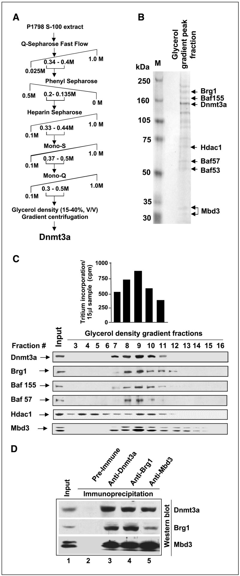
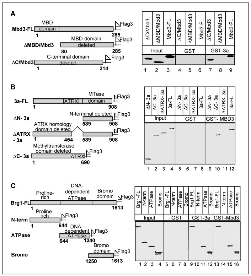
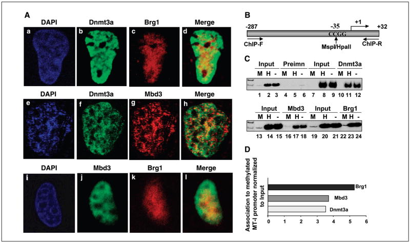
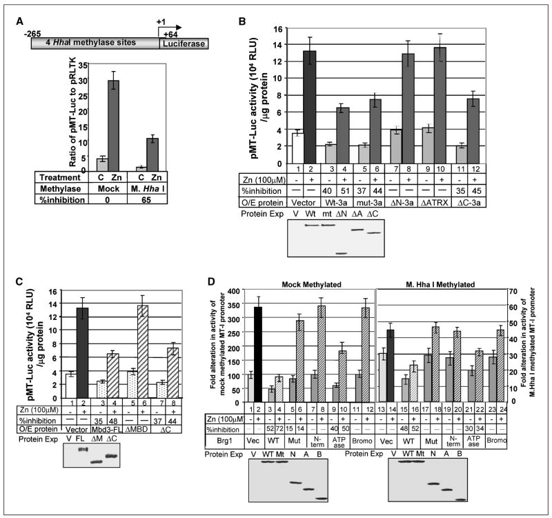
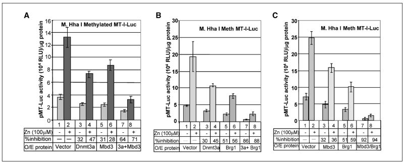
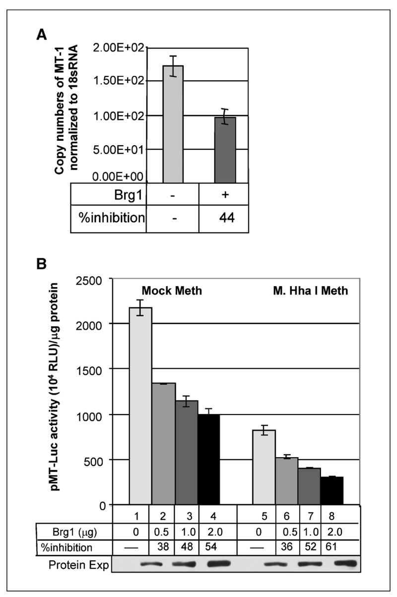
Dnmt3a and Dnmt3b are de novo DNA methyltransferases that also act as transcriptional repressors independent of methyltransferase activity. To elucidate the underlying mechanism of transcriptional repression, Dnmt3a was purified from mouse lymphosarcoma cells (P1798) by extensive fractionation on five different chromatographic matrices followed by glycerol density gradient centrifugation. Liquid chromatography electrospray tandem mass spectrometry analysis of Dnmt3a-associated polypeptides identified the methyl CpG binding protein Mbd3, histone deacetylase 1(Hdac1), and components of Brg1 complex (Brg1, Baf155, and Baf57) in the purified preparation. Association of Dnmt3a with Mbd3 and Brg1 was confirmed by coimmunoprecipitation and coimmunolocalization studies. Glutathione S-transferase pulldown assay showed that the NH2-terminal ATRX homology domain of Dnmt3a interacts with the methyl CpG binding domain of Mbd3 and with both bromo and ATPase domains of Brg1. Chromatin immunoprecipitation assay revealed that all three proteins are associated with transcriptionally silent methylated metallothionein (MT-I) promoter in the mouse lymphosarcoma cells. To understand the functional significance of their association with the promoter, their role on the MT-I promoter activity was analyzed by transient transfection assay. The results showed that Mbd3 and Dnmt3a specifically inhibited the methylated promoter, and the catalytic activity of Dnmt3a was dispensable for the suppression. In contrast, the wild-type but not the ATPase-inactive mutant of Brg1 suppressed MT-I promoter irrespective of its methylation status, implicating involvement of ATP-dependent chromatin remodeling in the process. Coexpression of two of the three interacting proteins at a time augmented their repressor function. This study shows physical and functional interaction of Dnmt3a with components of nucleosome remodeling machinery.


Figures

Figure 1
Figure 1
Profile of polypeptides copurified with Dnmt3a. A, fractionation scheme of Dnmt3a from mouse lymphosarcoma S-100 extract. B, identity of polypeptides copurified with Dnmt3a as determined by LC-MS/MS. The fraction of glycerol density gradient maximally enriched in Dnmt3a was separated on SDS-PAGE. Major polypeptides stained with Coomassie blue were subjected to MS. C, Western blot analysis of glycerol density gradient fractions with specific antibodies. Top, de novo DNA methyltransferase activity measured in glycerol density gradient fractions enriched in Dnmt3a. Bottom, location of Dnmt3a and other proteins of nucleosome remodeling complex. D, Dnmt3a coimmunoprecipitates with Brg1 and Mbd3. Mouse lymphosarcoma cell extracts (500 μg) were immunoprecipitated with anti-Dnmt3a, anti-Brg1, or anti-Mbd3 antibodies in TNN buffer. The corresponding preimmune serum served as the control. The immunoprecipitated polypeptides were analyzed by Western blotting sequentially with the antibodies against each protein after stripping in the buffer [100 mmol/L Tris-HCl (pH 6.8), 2%SDS, 100 mmol/L mercaptoethanol] at 55°C for 30 minutes.
Figure 2
Figure 2
ATRX homology domain of Dnmt3a, MBD of Mbd3, and both ATPase and bromo domains of Brg1 are involved in protein-protein interaction as shown by GST pulldown assay. A, schematic representation of full-length as well as different domains of Mbd3 fused to 3xFlag and/or GST. For pull down assay, GST alone or GST-Dnmt3a coupled to GSH-Sepharose beads were allowed to interact with HepG2 cell extract over expressing the wild type (Mbd3-FL-Flag), or different deletion mutants of Mbd3 under stringent condition (see Materials and Methods for details). The pulled down wild type and mutant Mbd3 were detected by Western blotting with anti-Flag antibody. B, schematic representation of full-length and different deletion mutants of Dnmt3a with COOH-terminal Flag tag. For pull down assay, GST alone or GST-Mbd3 coupled to GSH-Sepharose was allowed to bind to the wild type or different deletion mutants of Dnmt3a overexpressed in HepG2 cells. C, schematic diagram depicting full-length and different domain deletion mutants of Brg1 fused to 3xFlag peptide. GST pull down assay was done with GST alone, GST-Dnmt3a, or GST-Mbd3 from HepG2 cell extract overexpressing wild type and variants of Brg1-Flag (see Materials and Methods for additional details for AC).
Figure 3
Figure 3
Dnmt3a, Brg1, and Mbd3 colocalize in HepG2 cells. HepG2 cells were transiently transfected with expression vector for Dnmt3a, Mbd3, and Brg1 in different combination, fixed, and then immunostained as described in Materials and Methods. The cells expressing the recombinant proteins were visualized by confocal microscopy. Dnmt3a was visualized using affinity-purified rabbit polyclonal antibody raised against Dnmt3a, whereas Mbd3 and Brg1 were identified following overexpression as Flag-tagged protein and staining with anti-Flag antibody. A, when Mbd3 was coexpressed with Flag-tagged Brg1, affinity-purified rabbit polyclonal Mbd3 antibody was used to detect Mbd3. Colocalization of Dnmt3a and Brg1 (ad), Dnmt3a and Mbd3 (eh), and Mbd3 and Brg1 (il), respectively (see Materials and Methods for details). B, schematic depiction of MT-I promoter region amplified from the immunoprecipitated DNA. C, chromatin immunoprecipitation of MT-I promoter by anti-Dnmt3a, anti-Mbd3, and anti-Brg1 antibodies. Formaldehyde cross-linked chromatin from P1798 cells was immunoprecipitated with anti-Dnamt3a, Mbd3, or Brg1 antibodies (see Materials and Methods for details), and the precipitated DNA was divided into three aliquots that were digested with MspI, HpaII, or mock digested. MT-I promoter was amplified from these DNAs with specific primers. D, quantitative analysis of the association of these proteins with MT-I promoter. The amount of promoter amplified was quantitated using ImageQuant software and expressed as ratio of promoter amplified from immunoprecipitated DNA to that amplified from input DNA.
Figure 4
Figure 4
Modulation of MT-I promoter activity by Dnmt3a, Mbd3, and Brg1 in transient transfection assay. A, schematic diagram showing HhaI sites on mouse MT-I promoter. Methylation of these sites by M. HhaI methylase represses the constitutive (C) or Zn-induced (Zn) promoter activity. HepG2 cells were transfected with mock or methylated pMT-Luc along with pRL-tk. After 24 hours, cells were split into two and were either left untreated (−) 12 hours later or treated with 100 μmol/L zinc sulfate (+) for an additional 6 hours. The luciferase activity was measured in whole cell extract using Dual Luciferase assay kit (Promega, Madison, WI). B, Dnmt3a suppresses methylated MT-I promoter activity. HepG2 cells were cotransfected with the mammalian expression vector (Vector), wild-type Dnmt3a (Wt-3a), catalytic site mutant (mut-3a), and different deletion mutants (Fig. 2B) of the protein along with M. HhaI methylated pMT-Luc. Zn treatment and luciferase assay was done as described for (A). Western blot analysis of the overexpressing proteins (bottom). C, MBD of Mbd3 is required for suppressing methylated MT-I promoter activity. HepG2 cells were cotransfected with the mammalian expression vector (Vector), full-length Mbd3 (Mbd3-FL), MBD-deleted Mbd3 (ΔMBD), and COOH-terminal domain–deleted (ΔC) mutants (Fig. 2A) along with M. HhaI methylated pMT-Luc. Zn treatment and luciferase assay was done as described for (A). Western blot analysis of the overexpressing proteins (bottom). D, the wild type but not the ATPase domain mutant of Brg1 inhibits MT-I promoter activity irrespective of its methylation status. HepG2 cells were transfected with wild-type Brg1 (WT), catalytic site mutant (Mut), different domains of Brg1 (Fig. 2C), or empty vector (Vec) along with mock-methylated or M. HhaI–methylated pMT-I-Luc (see Materials and Methods for additional details). Luciferase activity in each sample (in arbitrary units) normalized to same amount of protein in each extract was compared and represented as fold alteration of the promoter activity upon overexpression of different Brg1 mutants. Western blot analysis of the overexpressing proteins (bottom).
Figure 5
Figure 5
Coexpression of Dnmt3a-associated proteins potentiates the repressive activity of Dnmt3a on methylated MT-I promoter. A, repressive effect of Mbd3 and Dnmt3a is additive when coexpressed. HepG2 cells were transfected with Dnmt3a and Mbd3 expression vectors alone or in combination, along with methylated pMT-Luc. After 24 hours, cells were split into two, and 12 hours later, these cells were either left untreated (−) or treated with 100 μmol/L zinc sulfate (+) for an additional 6 hours. The luciferase activity was measured in whole cell extract using Dual Luciferase assay kit (Promega). Luciferase activity in each sample (in arbitrary units) normalized to same amount of protein in each extract was compared. B, coexpression of Brg1 and Dnmt3a potentiates their repressive effect. Dnmt3a and Brg1 expression vectors along with methylated pMT-Luc were transfected alone or in combination into HepG2 cells. The cells were treated with Zn and luciferase activity measured as described (A). C, Brg1 and Mbd3 coexpression increases efficiency of silencing of methylated MT-I promoter. Mbd3 and Brg1 expression vectors were transfected alone or in combination into HepG2 cells along with methylated pMT-Luc. The cells were treated with Zn, and luciferase activity measured as described in (A).
Figure 6
Figure 6
Ectopic expression of Brg1 suppresses the MT-I promoter activity in Brg1 null (SW13) cells. A, MT-I expression is suppressed after ectopic expression of mBrg1 in SW13 cells. cDNAs prepared from SW13 cells after 36 hours of transfection with pBJ5 or pBJ5-Brg1 (2 μg) were subjected to real-time PCR with primers specific for human MT-I or 18S rRNA. Copy number for each mRNA was calculated based on a standard curve generated from 10-fold serial dilutions (108 to 102) of respective cDNAs. Each sample was assayed in triplicate. Columns, mean of three separate experiments; bars, SD. B, dose-dependent inhibition of MT-I promoter activity after ectopic expression of Brg1 in SW13 cells. SW13 cells were cotransfected with the mammalian expression vector (2 μg), Brg1 (0.5–2 μg) along with mock/M. HhaI–methylated pMT-Luc. Luciferase assay was done as described for Fig. 5A. Luciferase activity in each sample (in arbitrary units) normalized to same amount of protein in each extract was compared. Western blot analysis of the overexpressing Brg1 protein (bottom).

References

    1. Okano M, Xie S, Li E. Cloning and characterization of a family of novel mammalian DNA (cytosine-5) methyl-transferases. Nat Genet. 1998;19:219–20. - PubMed
    1. Okano M, Bell DW, Haber DA, Li E. DNA methyl-transferases Dnmt3a and Dnmt3b are essential for de novo methylation and mammalian development. Cell. 1999;99:247–57. - PubMed
    1. Hata K, Okano M, Lei H, Li E. Dnmt3L cooperates with the Dnmt3 family of de novo DNA methyltransferases to establish maternal imprints in mice. Development. 2002;129:1983–93. - PubMed
    1. Reik W, Walter J. Genomic imprinting: parental influence on the genome. Nat Rev Genet. 2001;2:21–32. - PubMed
    1. Li E. Chromatin modification and epigenetic reprogramming in mammalian development. Nat Rev Genet. 2002;3:662–73. - PubMed

Publication types

MeSH terms