Comparative Study
. 2006 Apr;290(4):R951-62.
doi: 10.1152/ajpregu.00734.2005. Epub 2005 Dec 1.

Role for pituitary adenylate cyclase activating polypeptide in cystitis-induced plasticity of micturition reflexes

Affiliations
Comparative Study

Role for pituitary adenylate cyclase activating polypeptide in cystitis-induced plasticity of micturition reflexes

Karen M Braas et al. Am J Physiol Regul Integr Comp Physiol. 2006 Apr.

Abstract

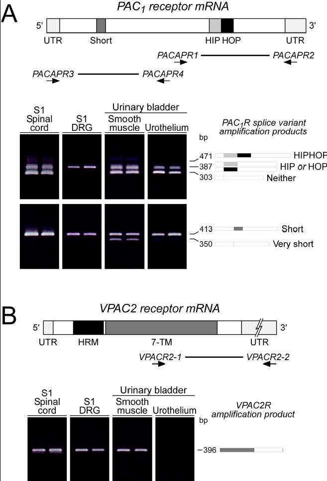
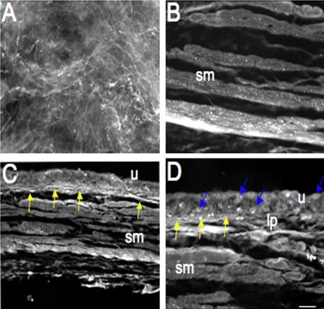
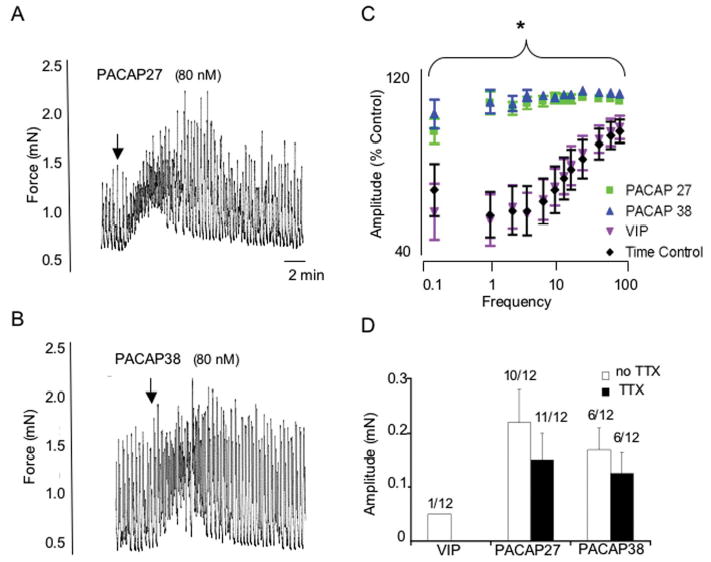
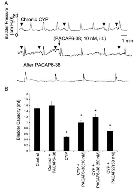
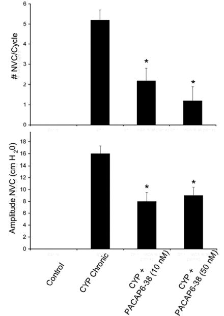
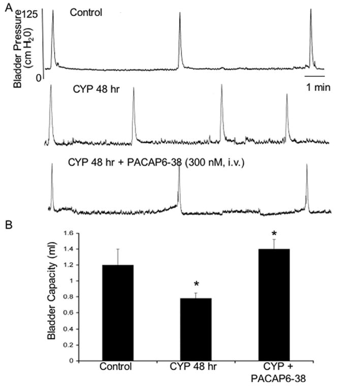
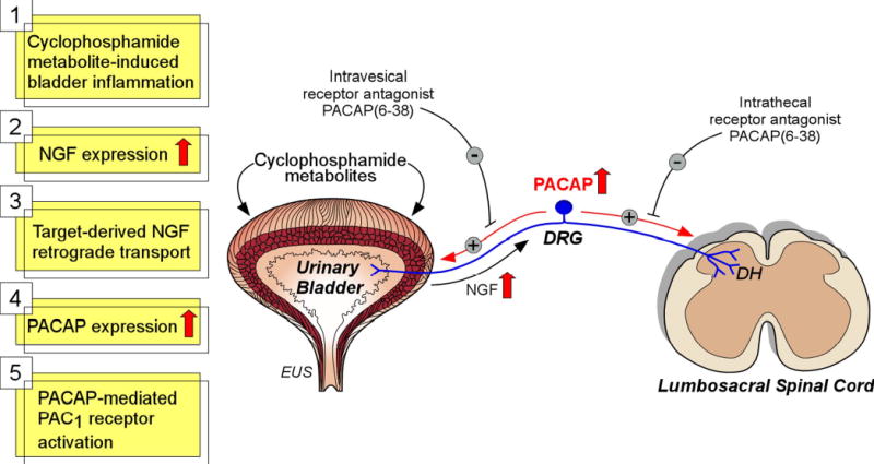
Pituitary adenylate cyclase activating polypeptide (PACAP) peptides are expressed and regulated in sensory afferents of the micturition pathway. Although these studies have implicated PACAP in bladder control, the physiological significance of these observations has not been firmly established. To clarify these issues, the roles of PACAP and PACAP signaling in micturition and cystitis were examined in receptor characterization and physiological assays. PACAP receptors were identified in various tissues of the micturition pathway, including bladder detrusor smooth muscle and urothelium. Bladder smooth muscle expressed heterogeneously PAC(1)null, PAC(1)HOP1, and VPAC(2) receptors; the urothelium was more restricted in expressing preferentially the PAC(1) receptor subtype only. Immunocytochemical studies for PAC(1) receptors were consistent with these tissue distributions. Furthermore, the addition of 50-100 nM PACAP27 or PACAP38 to isolated bladder strips elicited transient contractions and sustained increases in the amplitude of spontaneous phasic contractions. Treatment of the bladder strips with tetrodotoxin (1 muM) did not alter the spontaneous phasic contractions suggesting direct PACAP effects on bladder smooth muscle. PACAP also increased the amplitude of nerve-evoked contractions. By contrast, vasoactive intestinal polypeptide had no direct effects on bladder smooth muscle. In a rat cyclophosphamide (CYP)-induced cystitis paradigm, intrathecal or intravesical administration of PAC(1) receptor antagonist, PACAP6-38, reduced cystitis-induced bladder overactivity. In summary, these studies support roles for PACAP in micturition and suggest that inflammation-induced plasticity in PACAP expression in peripheral and central micturition pathways contribute to bladder dysfunction with cystitis.


Figures

Figure 1
Figure 1
A. Lower urinary tract (LUT) tissues express PAC1 receptor variants. Complementary DNA templates were prepared from rat S1 spinal cord, S1 dorsal root ganglia (DRG) and bladder detrusor and urothelium total RNA. The region spanning the alternative splice site for the HIP and HOP exons within the third cytoplasmic loop was amplified using PACAPR1/2 oligonucleotide primers. Six third cytoplasmic loop isoform fragments containing neither, one or both HIP and HOP cassettes can potentially be amplified with these primers. LUT tissues express PAC1 receptor isoforms in a tissue-specific manner. S1 DRG express predominantly the one cassette isoform; other tissues possess both the null and the one cassette variant. Schematic shading: Dark grey, short region containing exons 4 and 5; light grey, HIP exon cassette; black, HOP cassette. Thick line, region amplified using PACAPR1/2 primers. Lower urinary tract (LUT) tissue expression of PAC1 receptor isoforms also result from alternative splicing in amino-terminal extracellular domain. Complementary DNA templates from LUT samples described above were amplified using primers PACAPR3/4 which flank the amino-terminal extracellular domain splice site. The amplified fragments of indicated sizes represent isoforms with both (short) or neither (very short) exons 4 and 5. All LUT tissues express the short variant; urinary detrusor smooth muscle also demonstrates very short PAC1 receptor expression. Shading in schematic denotes alternatively spliced exons. Thick line, region amplified using primers PACAR3/4. B. VPAC2 receptor expression in LUT tissues. cDNA from LUT tissues were prepared as described for amplification using oligonucleotide primers VPACR1/2 that span the carboxy-terminal domain of the 7 transmembrane (7-TM) receptor. All LUT tissues except bladder urothelium express VPACR2 receptor transcripts. Dark grey, 7 transmembrane domain; black, hormone receptor domain (HRM). Thick line, region amplified using primers VPACR1/2.
Figure 2
Figure 2
Fluorescence photographs of PAC1 receptor immunoreactivity (IR) in urinary bladder in control (A–D) rats. PAC1-IR was present in control bladder in the urothelium (u), suburothelial plexus (A, C, D, yellow arrows) and detrusor smooth muscle (B, sm). Individual urothelial cells express PAC1-IR (D, blue arrows). PAC1-IR in the suburothelial plexus was also examined in whole mount preparations (A). Calibration bar represents 100 μm in A and 80 μm in B–D.
Figure 3
Figure 3
PACAP27 (80 nM; A) and PACAP 38 (80 nM; B) increased detrusor smooth muscle tone whereas VIP only rarely resulted in changes in tone (D). These changes in detrusor tone were not blocked by tetrodotoxin (1 μM) suggesting a direct effect of PACAP on detrusor smooth muscle (D). PACAP27 or PACAP 38 (80 nM) potentiated the amplitude of nerve-evoked contractions at all frequencies tested (C) whereas no difference in electric field stimulation induced contraction amplitude was observed between time control and VIP-treated detrusor strips (C). *, p ≤ 0.001.
Figure 4
Figure 4
Intrathecal (i.t.) administration of PAC1 receptor antagonist, PACAP6-38 (10 nM) reduces voiding frequency (increased bladder capacity) after chronic cyclophosphamide (CYP)-induced cystitis (A). A. Continuous cystometrogram recording from the same rat treated chronically with CYP and then administered PACAP6-38 (10 nM; i.t.) with recording continuing after drug treatment. Intrathecal PACAP6-38 also reduced the number and amplitude of nonvoiding bladder contractions (NVCs) induced after CYP treatment. B. Summary histogram of bladder capacity in control, CYP-treated or CYP-treated with PACAP6-38 (i.t., 10 or 50 nM). CYP treatment significantly (p ≤ 0.001) reduced bladder capacity but this was significantly (p ≤ 0.001) increased after i.t. PACAP6-38 (10 or 50 nM) and again reduced after i.t. administration of agonist, PACAP27 (50 nM). PACAP6-38 (i.t., 10 or 50 nM) was without effect on bladder capacity in control rats.
Figure 5
Figure 5
A. Summary histogram of non-voiding bladder contractions (NVCs) induced by chronic cyclophosphamide (CYP) treatment that were significantly (*, p ≤ 0.001) reduced with intrathecal PACAP6-38 (10 nM and 50 nM). B. Summary histogram of amplitude of NVCs induced by chronic CYP treatment that were significantly (*, p ≤ 0.001) reduced in amplitude with intrathecal PACAP6-38 (10 nM and 50 nM) treatment.
Figure 6
Figure 6
A. Bladder pressure recordings in control rats and in those treated with cyclophosphamide (CYP; 48 hr). CYP treatment (48 hr) significantly (p ≤ 0.01) decreased bladder capacity (increased voiding frequency) (A). Intravesical PACAP6-38 (300 nM) significantly (p ≤ 0.01) increased bladder capacity compared to the CYP treatment group alone (A, B). i.v., intravesical.
Figure 7
Figure 7
Potential mechanism underlying PACAP’s role in detrusor overactivity with CYP-induced bladder inflammation. Previous studies have demonstrated increases in nerve growth factor (NGF) in the urinary bladder with CYP-induced cystitis (25, 46, 67). NGF is retrogradely transported from the urinary bladder to dorsal root ganglia (DRG). NGF can upregulate PACAP expression in DRG and spinal cord with CYP-induced cystitis (69). The PAC1 receptor antagonist, PACAP6-38, was effective in reducing bladder overactivity with both intrathecal and intravesical administration. PACAP6-38 may act at the urinary bladder, DRG and spinal cord to reduce detrusor overactivity induced by CYP. MPG, major pelvic ganglion; DH, dorsal horn; SPN, sacral parasympathetic nucleus; INT, interneurons; PGN, preganglionic neurons; EUS, external urethral sphincter.

References

    1. Arimura A. Perspectives on pituitary adenylate cyclase activating polypeptide (PACAP) in the neuroendocrine, endocrine, and nervous systems. Jpn J Physiol. 1998;48:301–331. - PubMed
    1. Arimura A, Somogyvari-Vigh A, Miyata A, Mizuno K, Coy DH, Kitada C. Tissue distribution of PACAP as determined by RIA: highly abundant in the rat brain and testes. Endocrinology. 1991;129:2787–2789. - PubMed
    1. Beaudet MM, Parsons RL, Braas KM, May V. Mechanisms mediating pituitary adenylate cyclase-activating polypeptide depolarization of rat sympathetic neurons. J Neurosci. 2000;20:7353–7361. - PMC - PubMed
    1. Birder LA, Ruan HZ, Chopra B, Xiang Z, Barrick S, Buffington CA, Roppolo JR, Ford AP, de Groat WC, Burnstock G. Alterations in P2X and P2Y purinergic receptor expression in urinary bladder from normal cats and cats with interstitial cystitis. Am J Physiol Renal Physiol. 2004;287:F1084–1091. - PubMed
    1. Braas KM, May V. Pituitary adenylate cyclase-activating polypeptides directly stimulate sympathetic neuron neuropeptide Y release through PAC(1) receptor isoform activation of specific intracellular signaling pathways. J Biol Chem. 1999;274:27702–27710. - PubMed

Publication types

MeSH terms

Substances

LinkOut - more resources