. 2006 Apr 1;107(7):2653-61.
doi: 10.1182/blood-2005-10-4035. Epub 2005 Dec 1.

Self-complementary adeno-associated virus vectors containing a novel liver-specific human factor IX expression cassette enable highly efficient transduction of murine and nonhuman primate liver

Affiliations

Self-complementary adeno-associated virus vectors containing a novel liver-specific human factor IX expression cassette enable highly efficient transduction of murine and nonhuman primate liver

Amit C Nathwani et al. Blood. .

Abstract

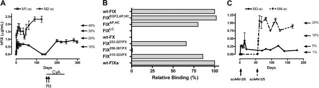
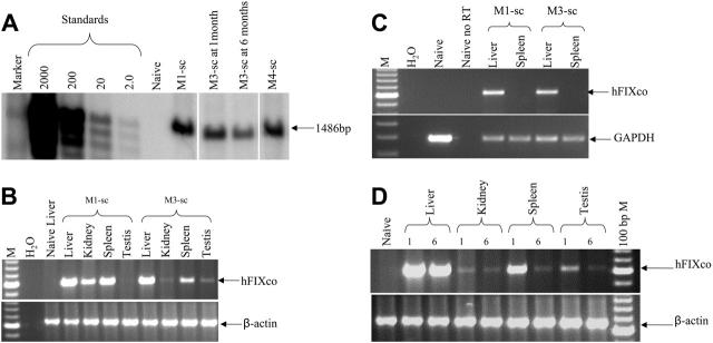
Transduction with recombinant adeno-associated virus (AAV) vectors is limited by the need to convert its single-stranded (ss) genome to transcriptionally active double-stranded (ds) forms. For AAV-mediated hemophilia B (HB) gene therapy, we have overcome this obstacle by constructing a liver-restricted mini-human factor IX (hFIX) expression cassette that can be packaged as complementary dimers within individual AAV particles. Molecular analysis of murine liver transduced with these self-complementary (sc) vectors demonstrated rapid formation of active ds-linear genomes that persisted stably as concatamers or monomeric circles. This unique property resulted in a 20-fold improvement in hFIX expression in mice over comparable ssAAV vectors. Administration of only 1 x 10(10) scAAV particles led to expression of hFIX at supraphysiologic levels (8I U/mL) and correction of the bleeding diathesis in FIX knock-out mice. Of importance, therapeutic levels of hFIX (3%-30% of normal) were achieved in nonhuman primates using a significantly lower dose of scAAV than required with ssAAV. Furthermore, AAV5-pseudotyped scAAV vectors mediated successful transduction in macaques with pre-existing immunity to AAV8. Hence, this novel vector represents an important advance for hemophilia B gene therapy.


Figures

Figure 1.
Figure 1.
Construction and characterization of scAAV vectors. (A) Structure of rAAV hFIX vectors. Each vector is shown schematically as it is packaged in the virion, with scAAV vectors shown as dimers. ssAAV-HCR-hAAT-FIX consists of the human apolipoprotein E/C-I gene locus control region (HCR) and the human α1 antitrypsin promoter (hAAT), a chicken β actin/rabbit β globin composite intron (IVS), 1.6-kb human FIX cDNA (hFIX), and a bovine growth hormone polyadenylation signal (BGpA) flanked by the AAV internal terminal repeats (ITRs shown as hairpin loop). Self-complementary scAAV-LP1-hFIXco vector containing the LP1 promoter consisting of core liver-specific elements from HCR (base pairs 134 to 442 of GenBank record HSU32510) and hAAT (base pairs 1747 to 2001 of GenBank record K02212), modified SV40 small t antigen intron (base pairs 4644 to 4552 of GenBank record J02400), codon-optimized hFIX (hFIXco), and SV40 late polyA (SV40 LpA; base pairs 2600 to 2733 of GenBank record J02400), and a deleted 3′ trstrs). scAAV-LP1-hFIX contains the wild-type human FIX cDNA from which the 3′ untranslated region has been deleted (ΔhFIX) instead of hFIXco. ssAAV-LP1-hFIXcoS has intact 5′ and 3′ ITR and in addition contains 760 bp noncoding stuffer sequence from the β-lactamase (Δbla). (B) Characterization of ss and scAAV2/8 viral particles. Viral particles were electrophoresed on a 1% neutral or alkaline agarose gel, Southern blotted, and hybridized with a vector-specific probe. The controls consisted of approximately 4.6- and 2.3-kbp fragments that were derived from ssAAV HCR-hAAT FIX and scAAV LP1-hFIXco plasmids cut with a double cutter, which cuts outside the ITR regions. The scAAV genome migrated with the 4.6-kb marker on the alkaline agarose gel and 2.3-kb marker on neutral gel, while the migration pattern of ssAAV vectors of differing size on these 2 gels did not change.
Figure 2.
Figure 2.
Influence of genomic configuration and capsid proteins on transduction efficiency. (A) Plasma hFIX levels at 6 weeks after tail-vein administration of varying doses of ssAAV2/8-HCR-hAAT-FIX (▵, n = 4/dose) and scAAV2/8-LP1-hFIXco (□, n = 4/dose). Results are depicted as average together with the standard error of the mean (error bars are not visible at some time points because of the log scale). (B) Representative results of immunohistochemical staining for hFIX at 7 days and 42 days (middle 2 panels) after tail-vein administration of 1 × 10¹¹ scAAV2/8-LP1-hFIXco compared with naive liver (far-left panel) from a control animal. The far-right panel shows hFIX expression in hepatocytes at 42 days after tail-vein administration of 1 × 10¹¹ ssAAV2/8 vector. Magnification is × 40. (C) Influence of AAV capsid proteins on scAAV-LP1-hFIXco–mediated-hFIX expression. Plasma hFIX levels assessed at 4 weeks after tail-vein administration of 1 × 10¹¹ vg of either scAAV2/8-LP1-hFIXco (n = 4) or scAAV2/5-LP1-hFIXco (n = 4). Results are depicted as the average together with the standard error of the mean.
Figure 3.
Figure 3.
Low-dose transduction efficiency with ss and scAAV vectors in murine models. (A) Transgene expression in C57Bl/6 mice. hFIX-expression profile over 31 weeks after tail-vein administration of 2 × 10⁹ vg/mouse (n = 3) of scAAV2/8-LP1-hFIXco (▵), scAAV2/8-LP1-hFIX (□), ssAAV2/8-LP1-hFIXcoS (○), and ssAAV2/8-HCR-hAAT-FIX (⋄) into C57Bl/6 mice. All hFIX results are depicted as the average together with the standard error of the mean. (B) Correction of clotting times and expression of hFIX:C in 129/sv HB mice. Following tail-vein injection of either 1 × 10¹⁰ (low-dose cohort, n = 6) or 5 × 10¹⁰ (high-dose cohort, n = 4) scAAV2/8-LP1-hFIXco vector particles into 129/sv HB mice, the clotting time (left-hand panel at 4 weeks) and biologically active hFIX:C levels (right-hand panel) over 16 weeks were determined.
Figure 4.
Figure 4.
Molecular configuration of scAAV genome in murine liver. (A) Southern analysis of modified Hirt DNA extracted from the liver of a subset of mice at day 1 or 42. Approximately 15 μg DNA from each time point was electrophoresed uncut (uncut), or following digestion with single cutters (PstI) or plasmid safe DNase (PS-D). Shown are bands representing monomeric supercoiled circular (MSC) or dimeric supercoiled circular (DSC), ds-linear (L-ds), or high-molecular-weight concatamers (HMWCs) in the head-tail (HT) and head-to-head (HH) formation. For comparison, 5 × 10⁶ and 2 × 10⁷ purified vector particles were loaded directly on the gel after denaturation (last 2 lanes). Bottom panel is a schematic representation of dimeric scAAV-LP1-hFIXco in the HT (top) and HH (bottom) configuration together with the sites of cleavage by PstI. (B) Southern blot analysis of liver DNA isolated before and after partial hepatectomy at 16 and 20 weeks, respectively, after tail-vein administration of 1 × 10¹⁰ scAAV2/8-LP1-hFIXco vector particles into 129/sv HB mice (HBM1 and 2). Each lane contains 10 μg DNA digested with EcoRI and PstI, which releases a 1.1-kb fragment. Proviral copy number (shown at the bottom) was deduced from standards, which consisted of serial dilutions of vector DNA (0.13 to 13 copies) in 10 μg negative genomic DNA.
Figure 5.
Figure 5.
Biodistribution of scAAV2/8-LP1-hFIXco following tail-vein administration of vector. (A) PCR analysis of 1 μg genomic DNA, isolated from the indicated organs 6 weeks after tail-vein (TV) administration of 1 × 10¹¹ particles using primers unique to hFIXco designed to amplify a 617-bp product. Control consists of genomic DNA extracted from the liver of an untransduced animal. scAAV2/8-LP1-hFIXco vector particles (3 × 10¹⁰) were also administered directly into the quadriceps muscle for comparison. Genomic DNA (1 μg) extracted from the muscle tissue was subjected to PCR using identical conditions. Integrity of DNA was determined by amplifying a 604-bp region of the murine β-actin gene and is shown at the bottom panel. (B) RT-PCR analysis. Expression analysis of hFIXco mRNA by RT-PCR following tail-vein injection of 1 × 10¹¹ or intramuscular administration of 3 × 10¹⁰ scAAV-LP1-hFIXco particles or mock-transduced mice (Naive liver). RNA samples were amplified with and without RT to exclude genomic DNA amplification. Integrity of the RNA was determined by amplifying a 295-bp region of the murine GAPDH gene and is shown at the bottom panel.
Figure 6.
Figure 6.
Transduction in rhesus macaques following liver-targeted delivery of scAAV2/8 and scAAV2/5-LP1-hFIXco. (A) Human FIX concentration in rhesus plasma was determined at the indicated time points after administration of 1 × 10¹² vg/kg (M1-sc [□], M2-sc [○]) scAAV2/8-LP1-hFIXco into the mesenteric vein of 2 rhesus macaques. Treatment of M1-sc with rituximab (Rit × 2 doses) and oral cyclosporine (CyA) is shown. (B) Graphic representation of the reactivity profile of the anti-hFIX antibody in M1-sc, as determined using a panel of hFIX/X chimeras with domain or surface loop substitutions derived from FX. Antibody binding to chimeras was expressed as percentage of binding to wild-type FIX. (C) Transgene expression after mesenteric-vein administration of 4 × 10¹¹ vg/kg scAAV2/8-LP1-hFIXco in 2 macaques (M3-sc [⋄] and M4-sc [▿]). M4-sc additionally received 1 × 10¹² vg/kg scAAV2/5-LP1-hFIXco at 56 days after initial exposure to scAAV2/8. Each sample was independently evaluated on at least 3 separate occasions, and the results are depicted as an average together with the standard error of the mean.
Figure 7.
Figure 7.
Molecular analysis in rhesus macaques. (A) Southern blot analysis of DNA (10 μg) derived from liver biopsy specimens from monkeys M1-sc and M4-sc 1 month after liver-targeted delivery of 1 × 10¹² vg/kg scAAV2/8-LP1-hFIXco and scAAV2/5-LP1-hFIXco, respectively, or at 1 and 6 months after administration of 4 × 10¹¹ vg/kg scAAV2/8-LP1-hFIXco in M3-sc. After digestion with BsrDI, the released 1486-bp fragment was probed with an LP1-specific probe. (B) Limited biodistribution analysis. Genomic DNA (top panel) isolated at 1 month from the indicated organs following liver-targeted administration of either 1 × 10¹² vg/kg or 4 × 10¹¹ vg/kg scAAV2/8-LP1-hFIXco into monkeys M1-sc or M3-sc, respectively, was subjected to PCR amplification using primers unique to hFIXco designed to amplify a 617-bp product. Integrity of DNA was determined by amplifying a 604-bp region of the rhesus β-actin gene and is shown at the bottom of this panel. (C) RT-PCR analysis of RNA extracted from the indicated organs following mesenteric-vein injection of either 1 × 10¹² vg/kg or 4 × 10¹¹ vg/kg scAAV2/8-LP1-hFIXco into monkeys M1-sc and M3-sc, respectively. RNA samples were amplified with (+) and without (–) RT to exclude genomic DNA amplification. Integrity of the RNA was determined by amplifying a 295-bp region of the rhesus GAPDH gene and is shown at the bottom of the panel. (D) Persistence of the scAAV transgene was assessed by subjecting genomic DNA (1 μg) isolated at 1 or 6 months from the indicated organs following liver-targeted administration of 4 × 10¹¹ vg/kg scAAV2/8-LP1-hFIXco in monkey M3-sc to PCR using primers unique to hFIXco designed to amplify a 617-bp product. Integrity of DNA was determined by amplifying a 604-bp region of the rhesus β-actin gene and is shown at the bottom of the panel.

Comment in

References

    1. Nathwani AC, Tuddenham EG. Epidemiology of coagulation disorders. Baillieres Clin Haematol. 1992;5: 383-439. - PubMed
    1. Nathwani AC, Davidoff AM, Tuddenham EG. Prospects for gene therapy of haemophilia. Haemophilia. 2004;10: 309-318. - PubMed
    1. Fisher KJ, Gao GP, Weitzman MD, et al. Transduction with recombinant adeno-associated virus for gene therapy is limited by leading-strand synthesis. J Virol. 1996;70: 520-532. - PMC - PubMed
    1. Nakai H, Storm TA, Kay MA. Annealing of complimentary single stranded genomes and subsequent intermolecular joining is the mechanism of stable in vivo liver transduction by recombinant adeno-associated virus vectors [abstract]. Mol Ther. 2000;1: S125-S126.
    1. Weitzman MD, Fisher KJ, Wilson JM. Recruitment of wild-type and recombinant adeno-associated virus into adenovirus replication centers. J Virol. 1996;70: 1845-1854. - PMC - PubMed

Publication types