

Skin-derived interleukin-7 contributes to the proliferation of lymphocytes in cutaneous T-cell lymphoma
- PMID: 16322477
- PMCID: PMC1895734
- DOI: 10.1182/blood-2005-03-1139
Skin-derived interleukin-7 contributes to the proliferation of lymphocytes in cutaneous T-cell lymphoma
Abstract
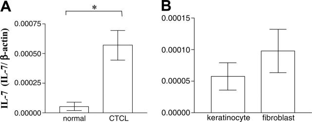
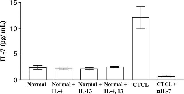
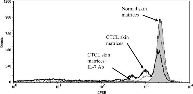
Cutaneous T-cell lymphomas (CTCLs) are malignancies of T cells that have a special affinity for the skin. We have previously reported that much of the T-cell receptor repertoire is altered in CTCL, and both malignant and nonmalignant clones are numerically expanded, presumably in response to T-cell trophic cytokines. We therefore examined levels of the T-cell trophic cytokines IL-2, IL-4, IL-7, IL-12, IL-13, and IL-15 in plasma in 93 CTCL patients and healthy controls. Only IL-7 levels were elevated in CTCL. We next looked at lesional skin from patients with CTCL and found elevated levels of IL-7 mRNA. Explant cultures of normal and lesional CTCL skin biopsies revealed significantly more IL-7 protein production in CTCL skin. Additionally, cultures of CTCL skin released greater numbers of T cells than normal skin; this was blocked by the addition of an IL-7 neutralizing antibody. Finally, these cultures induced proliferation of normal peripheral skin-homing T cells that were added to the cultures. These observations led us to postulate that IL-7 produced by skin cells contributes to the survival and proliferation of T cells within skin lesions and is likely the source of elevated circulating IL-7 in CTCL.
Figures


Similar articles
-
Expression of interleukin-18 and caspase-1 in cutaneous T-cell lymphoma.Clin Cancer Res. 2006 Jan 15;12(2):376-82. doi: 10.1158/1078-0432.CCR-05-1777. Clin Cancer Res. 2006. PMID: 16428475
-
Increased HMGB1 levels in lesional skin and sera in patients with cutaneous T-cell lymphoma.Eur J Dermatol. 2018 Oct 1;28(5):621-627. doi: 10.1684/ejd.2018.3400. Eur J Dermatol. 2018. PMID: 30378541
-
IL-22, but not IL-17, dominant environment in cutaneous T-cell lymphoma.Clin Cancer Res. 2011 Dec 15;17(24):7529-38. doi: 10.1158/1078-0432.CCR-11-1192. Epub 2011 Nov 2. Clin Cancer Res. 2011. PMID: 22048239
-
Cutaneous T cell lymphoma: the helping hand of dendritic cells.Ann N Y Acad Sci. 2001 Sep;941:1-11. Ann N Y Acad Sci. 2001. PMID: 11594563 Review.
-
Pathogenesis of cutaneous T-cell lymphoma: implications for the use of recombinant cytokines and photopheresis.Clin Exp Immunol. 1997 Jan;107 Suppl 1:16-20. Clin Exp Immunol. 1997. PMID: 9020930 Review.
Cited by
-
Interleukin-7 up-regulates cyclin D1 via activator protein-1 to promote proliferation of cell in lung cancer.Cancer Immunol Immunother. 2012 Jan;61(1):79-88. doi: 10.1007/s00262-011-1078-3. Epub 2011 Aug 17. Cancer Immunol Immunother. 2012. PMID: 21847632 Free PMC article.
-
Restoration of peripheral blood T cell repertoire complexity during remission in advanced cutaneous T cell lymphoma.Arch Dermatol Res. 2010 Aug;302(6):453-9. doi: 10.1007/s00403-009-1023-x. Epub 2010 Jan 29. Arch Dermatol Res. 2010. PMID: 20111968 Free PMC article.
-
The major isoforms of Bim contribute to distinct biological activities that govern the processes of autophagy and apoptosis in interleukin-7 dependent lymphocytes.Biochim Biophys Acta. 2012 Oct;1823(10):1877-93. doi: 10.1016/j.bbamcr.2012.06.017. Epub 2012 Jun 21. Biochim Biophys Acta. 2012. PMID: 22728771 Free PMC article.
-
Pediatric Burn Survivors Have Long-Term Immune Dysfunction With Diminished Vaccine Response.Front Immunol. 2020 Jul 21;11:1481. doi: 10.3389/fimmu.2020.01481. eCollection 2020. Front Immunol. 2020. PMID: 32793203 Free PMC article.
-
Mechanism, Consequences, and Therapeutic Targeting of Abnormal IL15 Signaling in Cutaneous T-cell Lymphoma.Cancer Discov. 2016 Sep;6(9):986-1005. doi: 10.1158/2159-8290.CD-15-1297. Epub 2016 Jul 15. Cancer Discov. 2016. PMID: 27422033 Free PMC article.
References
-
- Qin JZ, Zhang CL, Kamarashev J, Dummer R, Burg G, Dobbeling U. Interleukin-7 and interleukin-15 regulate the expression of the bcl-2 and c-myb genes in cutaneous T-cell lymphoma cells. Blood. 2001;98: 2778-2783. - PubMed
-
- Robert C, Kupper TS. Inflammatory skin diseases, T cells, and immune surveillance. N Engl J Med. 1999;341: 1817-1828. - PubMed
-
- Ferenczi K, Fuhlbrigge RC, Pinkus J, Pinkus GS, Kupper TS. Increased CCR4 expression in cutaneous T cell lymphoma. J Invest Dermatol. 2002;119: 1405-1410. - PubMed
-
- Uccini S, Ruco LP, Monardo F, La Parola IL, Cerimele D, Baroni CD. Molecular mechanisms involved in intraepithelial lymphocyte migration: a comparative study in skin and tonsil. J Pathol. 1993;169: 413-419. - PubMed
Publication types
MeSH terms
Substances
Grants and funding
LinkOut - more resources
Full Text Sources
Other Literature Sources
