Comparative Study
. 2006 Feb;172(2):811-25.
doi: 10.1534/genetics.105.050906. Epub 2005 Dec 1.

zds1, a novel gene encoding an ortholog of Zds1 and Zds2, controls sexual differentiation, cell wall integrity and cell morphology in fission yeast

Affiliations
Comparative Study

zds1, a novel gene encoding an ortholog of Zds1 and Zds2, controls sexual differentiation, cell wall integrity and cell morphology in fission yeast

Miyo Yakura et al. Genetics. 2006 Feb.

Abstract

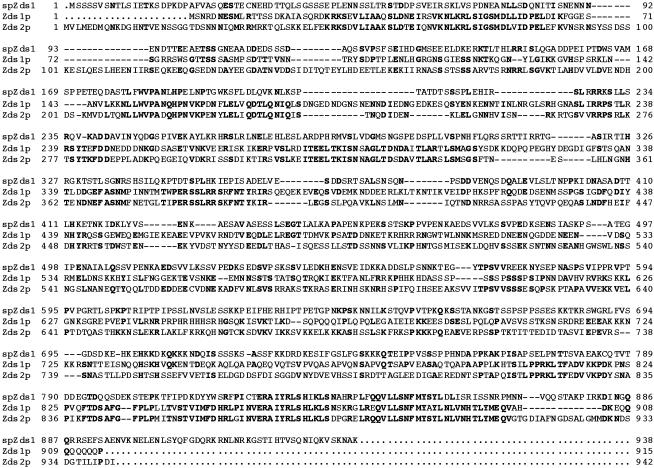
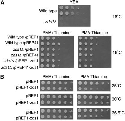
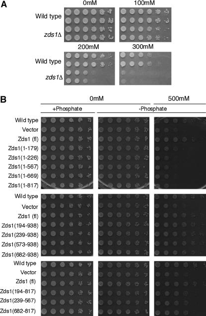
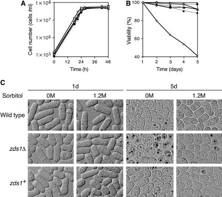
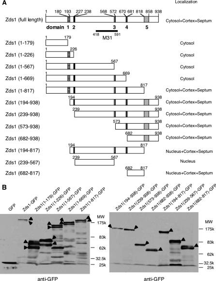
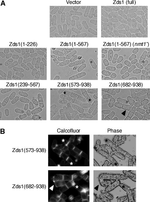
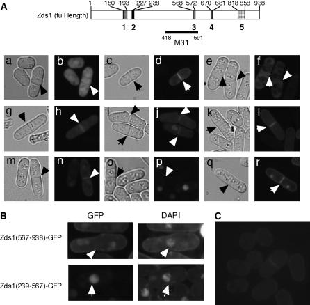
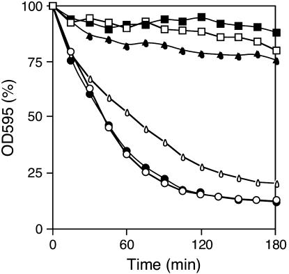
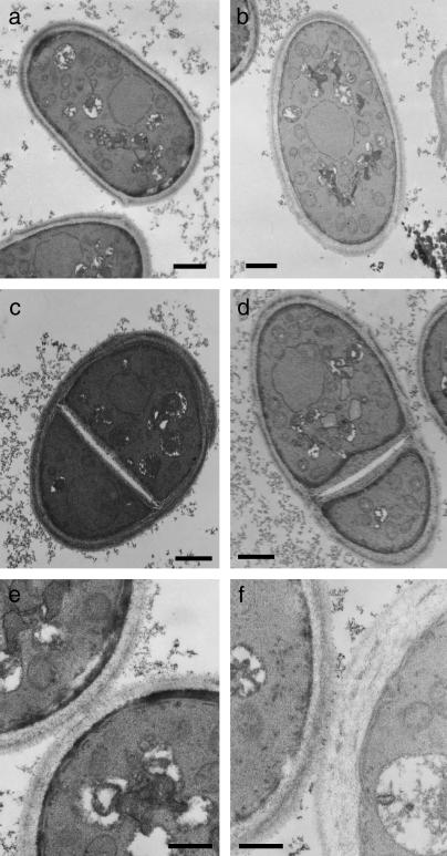
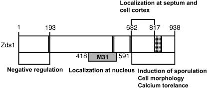
While screening for genes that reverse the sporulation-deficient phenotype of the ras1delta diploid Schizosaccharomyces pombe strain, we identified zds1. This gene shares sequence homology with the ZDS1 and ZDS2 genes from Saccharomyces cerevisiae, which appear to be involved in multiple cellular events. Expression of Zds1 in ras1delta diploid cells elevated their sporulation rate from 0.3 to 11.2%. Expression of the Zds1 C-terminal region increased the sporulation rate further (to 21.9%) while introduction of the Zds1 N-terminal region had no effect. zds1 expression did not induce sporulation in strains with mutations in genes participating in the downstream MAP kinase cascade. The zds1-disrupted strain is sensitive to CaCl2, and this effect is suppressed by the C-terminal region of Zds1. The growth of the zds1delta strain is markedly inhibited by cold temperatures, while its viability decreased in the stationary phase. Moreover, the zds1delta strain is round in shape and very sensitive to zymolyase, and its cell wall becomes thicker than that of wild type. Thus, zds1 must be required to maintain cell wall integrity. The Zds1-GFP fusion protein localized to the cytosol, the septum, and the cell cortex. Its localization in the septum was dependent on its C-terminal region. Overexpression of the C-terminal region of Zds1 induced multi-septa and abnormal zygotes. We propose that the C-terminal region is the functional domain of Zds1 while the N-terminal region is a negative regulatory region. Thus, Zds1 is involved in multiple cellular events in fission yeast, including sexual differentiation, Ca2+ tolerance, cell wall integrity, viability in the stationary phase, and cell morphology.


Figures

Figure 1.
Figure 1.
Comparison of the amino acid sequence of S. pombe (Sp) Zds1 with those of the Zds1p and Zds2p from S. cerevisiae. The putative nuclear localization signal is indicated by the underlined letters. The consensus amino acids are indicated by boldface type.
Figure 2.
Figure 2.
Temperature sensitivity of zds1-disrupted and zds1-overexpressing cells. (A) Wild-type and zds1-disruptant strains or those harboring the indicated plasmids were grown for 14 days at 16° on the indicated plates and growth was observed. (B) Wild type S. pombe cells (SP66) that overexpress zds1 due to transformation with pREP1–zds1 were spotted onto PMA plates in the presence or absence of thiamine, absence of which induces the expression of zds1 from the plasmids. The cells were incubated at 25°, 30°, or 36.5° for 5 days and growth was observed.
Figure 3.
Figure 3.
Calcium sensitivity of zds1-disrupted cells. (A) SP66 (h90, wild type) and MY6010 (h90, zds1Δ) cells were cultured at 30° in liquid medium until they reached log phase. They were concentrated to 2 × 10⁷ cells/ml and then diluted sequentially fivefold (to the right direction). The cells were spotted on YEA plates containing 0–300 mm CaCl2 and incubated at 30° for 3 days (0 and 100 mm CaCl2), 4 days (200 mm CaCl2), or 7 days (300 mm CaCl2). (B) The C-terminal region of Zds1 suppresses the calcium sensitivity of zds1-disrupted cells. SP66/pSLF272L GFPS65A cells were spotted on the plate as a wild-type strain. All other strains were transformants of MY6010. All plasmids were derived from the vector pSLF272L GFPS65A. The transformants were cultured at 30° in liquid PMA medium containing thiamine until they reached log phase, after which they were washed to remove the thiamine and resuspended in H2O. The cell concentration was adjusted to 2 × 10⁷ cells/ml and the cells were diluted sequentially fivefold (to the right direction). Cells were spotted on PMA (+phosphate), PMA containing sodium acetate instead of sodium phosphate (−phosphate; middle), or PMA (−phosphate; right) containing 500 mm CaCl2. These plates were incubated at 30° for 5 days (without CaCl2) or for 8 days (with CaCl2).
Figure 4.
Figure 4.
Low viability of zds1Δ in the stationary phase is suppressed under high osmotic condition. SP66 (h90, wild type)/pREP41, MY6010 (h90, zds1Δ)/pREP41, and MY6010/pREP41–zds1 (zds1+) were inoculated at a concentration of 1 × 10⁵ cells/ml in PMA medium with or without 1.2 m sorbitol and were cultured at 30°. (A) Growth curve. Cell number was counted using the Cell Counter (Sysmex). (B) Viability in the stationary phase. Cells were cultured until the indicated days. Cells were stained with methylene blue and ∼1000 cells, including unstained cells (living cells) and stained cells (dead cells), were counted under a microscope. SP66/pREP41 (open square), MY6010/pREP41 (open circle), and MY6010/pREP41–zds1 (open triangle) were grown without sorbitol. SP66/pREP41 (solid square), MY6010/pREP41 (solid circle), and MY6010/pREP41–zds1 (solid triangle) were grown with 1.2 m sorbitol. (C) Cell morphology. Indicated cells were grown as in B for 1 day and 5 days and observed. Asterisks indicate dead cells.
Figure 5.
Figure 5.
(A) Domain analysis of Zds1. Domain 1 (180–193) contains a sequence that is homologous to a region in S. cerevisiae Zds1p and Zds2p, domain 2 (227–238) contains a putative NLS, domain 3 (568–572) contains a region that is homologous to one in Zds2p, domain 4 (670–681) contains a Ser-rich sequence, and domain 5 (818–858) contains a putative functional domain. The M31 region is reported to localize to the nucleus (Sawin and Nurse 1996). (B) Western blot analysis of various Zds1–GFP fusion proteins. SP66 (wild-type) cells were transformed with the various plasmids shown in A and cultured to midlog phase. The proteins were then extracted. Arrowheads show various Zds1–GFP fusion proteins.
Figure 6.
Figure 6.
The morphology of cells expressing part of Zds1. (A) Wild-type SP66 was transformed with various plasmids that contain various lengths of the zds1 gene. Each transformant was cultured in PMA medium at 30° for 24 hr. The strains tested were SP66 harboring pSLF172L GFPS65A, pSLF172L Zds1–GFP, pSLF172L Zds1(1–226)–GFP, pSLF172L Zds1(1–567)–GFP, pSLF272L Zds1(1–567)–GFP, pSLF172L Zds1(239–567)–GFP, pSLF172L Zds1(573–938)–GFP, and pSLF172L Zds1(682–938)–GFP. (B) MY6010 cells expressing Zds1(573–938) and Zds1(682–938) were stained with calcofluor white. Asterisks show multi-septated cells and arrowheads show the abnormal zygotes.
Figure 7.
Figure 7.
The localization of various Zds1–GFP fusion proteins. Cells were cultured in PMA containing thiamine at 30° until they reached the stationary phase. The cells (at a density of 1 × 10⁵ cells/ml) were then inoculated into PMA without thiamine and further cultures at 30°. After 18 hr, the cells were observed under a fluorescent microscope. (A) MY6010/pSLF172L GFPS65A (a and b), MY6010/pSLF172L Zds1–GFP (c and d), MY6010/pSLF172L Zds1(1–669)–GFP (e and f), MY6010/pSLF172L Zds1(1–817)–GFP (g and h), MY6010/pSLF272L Zds1(194–938)–GFP (i and j), MY6010/pSLF172L Zds1(239–938)–GFP (k and l), MY6010/pSLF172L Zds1(573–938)–GFP (m and n), MY6010/pSLF172L Zds1(239–567)–GFP (o and p), and MY6010/pSLF172L Zds1(682–817)–GFP (q and r). Phase-contrast microscopic photographs (a, c, e, g, i, k, m, o, and q) and fluorescent microscopic photographs (b, d, f, h, j, l, n, p, and r) were taken. (B) Localization of Zds1ΔNΔC–GFP in nuclei. Cells were stained with DAPI and observed under a fluorescent microscope. (C) Localization of the Zds1–GFP fusion protein in the zds1–GFP integrated strain, MY6013. Cells were cultured in YES at 30° until they reached the stationary phase. They were then cultured in PM plus adenine and uracil until the midlog phase, washed with H2O and then with nitrogen-free PM medium, and resuspended in nitrogen-free PM medium. After 6 hr, the cells were observed by fluorescent microscopy.
Figure 8.
Figure 8.
Susceptibility of the zds1Δ strain to zymolyase. Cells were cultured in PMA medium at 30° to a density of 1 × 10⁷ cells/ml and then harvested and resuspended in TE (pH 8.0). Cells were incubated with 0.1 mg/ml zymolyase 20T at 30° for the indicated time. SP66/pREP41 (open square), MY6010/pREP41 (open circle), and MY6010/pREP41–zds1 (open triangle) were grown in the presence of thiamine. SP66/pREP41 (solid square), MY6010/pREP41 (solid circle), and MY6010/pREP41-zds1 (solid triangle) were grown without thiamine.
Figure 9.
Figure 9.
Electron micrographs of zds1-disrupted cells. Wild-type (SP66) and zds1-disrupted cells were cultured in PMAL medium at 30° for 72 hr and observed by TEM. Wild-type cells (a, c, and e), zds1-disrupted cells (b, d, and f), and septating cells of wild-type (c and e) and zds1-disrupted cells (d and f) were observed. The cells were observed at a magnification of 5000 (a and b), 6500 (c and d), and 15,000 (e and f). Bars, 1 μm in a, b, c, and d; 500 nm in e and f.
Figure 10.
Figure 10.
Summary of the Zds1 domain analysis conducted in this study. The N-terminal region serves to inhibit the functional domain of Zds1, which is the C-terminal region. The latter domain induces sexual development and calcium tolerance. The role of the central region is obscure but, interestingly, its sole expression induces nuclear localization.

Similar articles

Cited by

References

    1. Alfa, C., P. Fantes, J. Hyams, M. McLeod and E. Warbrick, 1993. Experiments With Fission Yeast: A Laboratory Course Manual. Cold Spring Harbor Laboratory Press, Cold Spring Harbor, NY.
    1. Bandhakavi, S., R. O. McCann, D. E. Hanna and C. V. Glover, 2003. Genetic interactions among ZDS1,2, CDC37, and protein kinase CK2 in Saccharomyces cerevisiae. FEBS Lett. 554: 295–300. - PubMed
    1. Barr, M. M., H. Tu, L. Van Aelst and M. Wigler., 1996. Identification of Ste4 as a potential regulator of Byr2 in the sexual response pathway of Schizosaccharomyces pombe. Mol. Cell. Biol. 16: 5597–5603. - PMC - PubMed
    1. Basi, G., E. K. Schmid and Maundrell, 1993. TATA box mutations in the Schizosaccharomyces pombe nmt1 promoter affect transcription efficiency but not the transcription start point or thiamine repressibility. Gene 123: 131–136. - PubMed
    1. Bi, E., and J. R. Pringle, 1996. ZDS1 and ZDS2, genes whose products may regulate Cdc42p in Saccharomyces cerevisiae. Mol. Cell. Biol. 16: 5264–5275. - PMC - PubMed

Publication types

MeSH terms

Substances