. 2006 Mar;172(3):1633-42.
doi: 10.1534/genetics.105.042911. Epub 2005 Dec 1.

De novo GMP synthesis is required for axon guidance in Drosophila

Affiliations

De novo GMP synthesis is required for axon guidance in Drosophila

Hong Long et al. Genetics. 2006 Mar.

Abstract

Guanine nucleotides are key players in mediating growth-cone signaling during neural development. The supply of cellular guanine nucleotides in animals can be achieved via the de novo synthesis and salvage pathways. The de novo synthesis of guanine nucleotides is required for lymphocyte proliferation in animals. Whether the de novo synthesis pathway is essential for any other cellular processes, however, remains unknown. In a search for genes required for the establishment of neuronal connectivity in the fly visual system, we identify the burgundy (bur) gene as an essential player in photoreceptor axon guidance. The bur gene encodes the only GMP synthetase in Drosophila that catalyzes the final reaction of de novo GMP synthesis. Loss of bur causes severe defects in axonal fasciculation, retinotopy, and growth-cone morphology, but does not affect photoreceptor differentiation or retinal patterning. Similar defects were observed when the raspberry (ras) gene, encoding for inosine monophosphate dehydrogenase catalyzing the IMP-to-XMP conversion in GMP de novo synthesis, was mutated. Our study thus provides the first in vivo evidence to support an essential and specific role for de novo synthesis of guanine nucleotides in axon guidance.


Figures
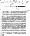
Figure 1.
Figure 1.
Molecular characterization of the bur gene. (A) Predicted genomic organization of the bur and two nearby genes mcm 10 and Ret at 39B1–2 by the BDGP/Celera Drosophila genome project. The exon–intron boundaries in the bur gene were determined by comparing the sequence of the full-length EST cDNA clone RE18382 to the genomic sequence. The P-element insertion site for burP is located within exon 1, 35 bp downstream of the putative bur transcriptional start site. In bure10, a C-to-T nonsense mutation in the third exon changes the codon for R61 into a stop codon. Open boxes, noncoding regions; solid boxes, coding regions. (B) Alignment of the Bur protein sequence with GMP synthetases in human (Hirst et al. 1994) and Escherichia coli (Tiedeman et al. 1985). Identical residues are boxed.
Figure 2.
Figure 2.
Mutations in the bur gene disrupted R-cell axon pathfinding. (A–C) R1–R8 axons in third-instar larvae were stained with mAb 24B10. In wild type (A), R-cell axons elaborate smooth retinotopic arrays in the lamina (la) and medulla (me). The expanded R1–R6 growth cones form a continuous layer in the lamina. R7 and R8 axons establish a smooth topographic array in the medulla, where individual expanded “Y-shaped” growth cones can be readily identified. (B) In a burP homozygote, thicker bundles (arrow) were frequently observed. Growth cones displayed irregular morphology. The array of R7 and R8 growth cones in the medulla was disorganized. (C) In a bure10 homozygote, the phenotype was much more severe. Axon bundles were much thicker than that in the burP mutant (B). Most R7 and R8 growth cones failed to expand and could not be identified individually. Crossing over of axons (arrow) was frequently observed. (D–F) Wild-type (D) or bur mutant axons (E and F) were labeled with the MARCM method (Lee and Luo 1999). (D) Labeled wild-type axons projected into appropriate topographic locations. Their growth cones expanded significantly when reaching the target region (arrow). (E) Labeled burP mutant axons in a mosaic larva did not expand their growth cones (arrow). (F) A labeled burP mutant axon turned abnormally along the dorsal–ventral axis and projected into an incorrect topographic location in medulla (arrow). Bar, 20 μm.
Figure 3.
Figure 3.
bur is not required for lamina-specific termination of R2–R5 axons. (A and B) R2–R5 axons in wild type (A) and bure10 mutant (B) were labeled with the larval R2–R5 marker ro-τ-lacZ. In wild type (A), the vast majority of ro-τ-lacZ-labeled R2–R5 axons terminated within the lamina. Only a few (i.e., 2–5) R2–R5 axons mistarget into the medulla. In a bure10 homozygote (B), the average number of mistargeted R2–R5 axons was 6 (n = 22 hemispheres), which was not significantly different from that in wild type (A). (C and D) R-cell axons (red) and laminal glial cells (green) in wild-type (C) and bure10 (D) third-instar larvae were double stained with anti-β-galactosidase antibody and anti-Repo antibodies, respectively. Both wild type and bure10 mutants carry a glass–lacZ transgene in which the expression of lacZ is under control of the eye-specific glass promoter (Mismer and Rubin 1987), which allows the visualization of all R-cell axons with anti-β-galactosidase staining. Anti-Repo recognizes a glial-specific nuclear protein. In wild type (C), glial cells (green) migrate from progenitor regions into the lamina where they are organized into two layers, the epithelial (eg) and marginal glia (mg), presenting a stop signal for the termination of R1–R6 growth cones (red) at the lamina plexus (lp). (D) In a bure10 homozygote, although glial cells (green) migrated correctly into the lamina, they appeared less organized. Bar, 20 μm.
Figure 4.
Figure 4.
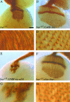
R-cell differentiation and patterning remained normal in bur mutants. (A and E) All R-cell bodies in the third-instar wild-type (A) and bure10 mutant (E) eye disc were visualized with mAb 24B10 staining. In wild type (A), ommatidial clusters of R-cell bodies are highly organized. (E) In a homozygous bure10 mutant eye disc, R-cell clusters maintained an appropriate neighbor relationship (n > 30 discs). (B and F) Differentiating R1–R8 nuclei in the third-instar wild-type (B) and bure10 mutant (F) eye disc were stained with anti-Elav antibody, which recognizes the panneuronal nuclear protein Elav. The organization of R-cell clusters in the bure10 mutant (F) (n = 44 discs) was indistinguishable from that in wild type (B). (C and G) R1 and R6 nuclei in the third-instar wild-type (C) and bure10 mutant (G) eye disc were stained with anti-Bar antibody. No obvious defect was observed in the bure10 mutant (n = 16 discs). (D and H) R8 cell bodies in the third-instar wild-type (D) and bure10 mutant (H) eye disc were stained with anti-Boss antibody. In a bure10 homozygote (H), like that in wild type (D), each ommatidium contains only a single R8 cell (n = 14 discs). Bar, 20 μm.
Figure 5.
Figure 5.
Rescue of the R-cell pathfinding phenotype by expressing the bur transgene in R cells. Third-instar eye–brain complexes were stained with mAb 24B10 to visualize R1–R8 axons. In a bure10 homozygote (A), R-cell axons displayed hyperfasciculation, aberrant topographic order, and growth-cone morphology. Genotype: bure10, GMR–GAL4/bure10. In a bure10 homozygote carrying a UASbur transgene under control of the eye-specific GMR–GAL4 driver (B), the R-cell projection pattern was indistinguishable from that in wild type (see Figure 2A). Note the appearance of expanded “Y-like” growth cones in the medulla. Genotype: bure10, GMR–GAL4/bure10; UASbur/+. (C and D) Enlarged views of the boxed regions in A and B, respectively. (E) In a bure10 homozygote expressing a UASmcm 10 transgene or a UAS–Ret transgene (data not shown), R-cell axons still displayed severe pathfinding defects. Genotype: bure10, GMR–GAL4/bure10; UASmcm 10/+. (F) In a bure10 homozygote carrying a UASbur transgene under control of the neuronal-specific elav–GAL4 driver, R-cell axons displayed a wild-type-like innervation pattern in the optic lobe. Genotype: elav–GAL4/+; bure10/bure10; UASbur/+. (G and H) Enlarged views of the boxed regions in E and F, respectively. Bars, 20 μm in A, B, E, and F; 5 μm in C, D, G, and H.
Figure 6.
Figure 6.
Mutations in the ras gene caused a bur-like phenotype in R-cell axon guidance. Third-instar eye–brain complexes were stained with mAb 24B10. (A) Wild type. (B) bure10 homozygote. (C) In a rasG0002 hemizygote, R-cell axons displayed a severe axon guidance phenotype (24 of 26 hemispheres examined) that was very similar to that in bure10. (D) A temperature-sensitive ras11 homozygote (100% penetrance, n = 92 hemispheres) grown at restrictive temperature (i.e., 29°). Bar, 20 μm.
Figure 7.
Figure 7.
Depleting guanine from the fly food did not affect R-cell axon guidance. Third-instar eye–brain complexes were stained with mAb 24B10. (A) R-cell projection pattern in a wild-type larva grown on normal food. (B) In a wild-type larva grown on guanine-depleted food, the R-cell projection pattern remained normal (n = 50 hemispheres). Bar, 20 μm.
Figure 8.
Figure 8.
Reducing the dosage of bur enhanced the Rac phenotype. Third-instar eye–brain complexes were stained with mAb 24B10. (A) R-cell projection pattern in a wild-type larva expressing the dominant-negative RacN17 transgene under control of the eye-specific GMR–GAL4 driver. The average number (25 ± 5, n = 12) of clearly separated axonal bundles between lamina and medulla was decreased compared to that in wild type (31 ± 6) due to axonal hyperfasciculation. Genotype: GMR–GAL4/+;UAS–RacN17/+;. (B) In a RacN17-expressing larva in which the dosage of bur was reduced by 50%, the phenotype became more severe. Compared to that in A (25 ± 5, n = 12), the average number (21 ± 4, n = 22) of axonal bundles was decreased (P < 0.05). Genotype: GMR–GAL4, bure10/+;UAS–RacN17/+. (C) In a larva expressing the dominant-negative RacL89 transgene, the R-cell projection pattern was severely disrupted. Genotype: GMR–GAL4/UAS–RacL89. (D) In a RacL89-expressing larva in which the dosage of bur was reduced by 50%, the R-cell axonal hyperfasciculation phenotype was enhanced. Compared to that in C (9 ± 5, n = 9), the average number (5 ± 2, n = 9) of axonal bundles was significantly decreased (P < 0.05). Genotype: GMR–GAL4, bure10/UAS–RacL89. Bar, 20 μm.

References

    1. Bork, P., and E. V. Koonin, 1994. A P-loop-like motif in a widespread ATP pyrophosphatase domain: implications for the evolution of sequence motifs and enzyme activity. Proteins 20: 347–355. - PubMed
    1. Carthew, R. W., and G. M. Rubin, 1990. seven in absentia, a gene required for specification of R7 cell fate in the Drosophila eye. Cell 63: 561–577. - PubMed
    1. Christensen, T. W., and B. K. Tye, 2003. Drosophila MCM10 interacts with members of the prereplication complex and is required for proper chromosome condensation. Mol. Biol. Cell 14: 2206–2215. - PMC - PubMed
    1. Clandinin, T. R., and S. L. Zipursky, 2002. Making connections in the fly visual system. Neuron 35: 827–841. - PubMed
    1. Dayton, J. S., L. A. Turka, C. B. Thompson and B. S. Mitchell, 1992. Comparison of the effects of mizoribine with those of azathioprine, 6-mercaptopurine, and mycophenolic acid on T lymphocyte proliferation and purine ribonucleotide metabolism. Mol. Pharmacol. 41: 671–676. - PubMed

Publication types

MeSH terms

Substances