. 2006 Jan;15(1):84-93.
doi: 10.1110/ps.051766506. Epub 2005 Dec 1.

Site-2 protease regulated intramembrane proteolysis: sequence homologs suggest an ancient signaling cascade

Affiliations

Site-2 protease regulated intramembrane proteolysis: sequence homologs suggest an ancient signaling cascade

Lisa N Kinch et al. Protein Sci. 2006 Jan.

Abstract

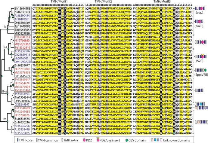
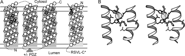
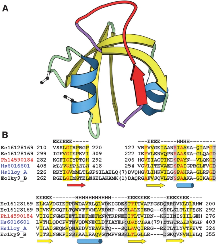
Site-2 proteases (S2Ps) form a large family of membrane-embedded metalloproteases that participate in cellular signaling pathways through sequential cleavage of membrane-tethered substrates. Using sequence similarity searches, we extend the S2P family to include remote homologs that help define a conserved structural core consisting of three predicted transmembrane helices with traditional metalloprotease functional motifs and a previously unrecognized motif (GxxxN/S/G). S2P relatives were identified in genomes from Bacteria, Archaea, and Eukaryota including protists, plants, fungi, and animals. The diverse S2P homologs divide into several groups that differ in various inserted domains and transmembrane helices. Mammalian S2P proteases belong to the major ubiquitous group and contain a PDZ domain. Sequence and structural analysis of the PDZ domain support its mediating the sequential cleavage of membrane-tethered substrates. Finally, conserved genomic neighborhoods of S2P homologs allow functional predictions for PDZ-containing transmembrane proteases in extra-cytoplasmic stress response and lipid metabolism.


Figures

Figure 1.
Figure 1.
Alignment and phylogeny of S2P core sequences from representative genomes. Sequences are labeled according to gi number, and species abbreviations are as follows: Bh, Bacillus halodurans; Tm, Thermotoga maritima; Tp, Treponema pallidum; Dr, Deinococcus radiodurans; Ss, Synechocystis sp.; Ml, Mycobacterium leprae; Tv, Thermoplasma volcanium; Hl, Halobacterium sp.; Mt, Methanothermobacter thermautotrophicus; Pa, Pyrobaculum aerophilum; At, Arabidopsis thaliana; Ce, Caenorhabditis elegans; Hs, Homo sapiens; Dm, Drosophila melanogaster; Pf, Plasmodium falciparum; Gl, Giardia lamblia. Sequence labels are colored according to superkingdom (bacteria, black; archaea, red; eukaryota, blue). TMH predictions by HMMTOP (Tusnady and Simon 2001) are shown above the alignment (H, TMH; i, intracellular; o, extracellular) with TMH motifs labeled above. Residues are highlighted according to chemical property conservation (hydrophobic, yellow; small, gray; charged/polar, black) and italicized (in Hl15790880 and At15235376) to mark omitted sequence insertions. In the unrooted tree to the left of the alignment, green circles denote internal tree nodes with at least 70% local bootstrap support. Reliably identified subfamilies are outlined with boxes, with the three ubiquitous ones colored gray. Four major groups of S2P subfamilies are labeled using Roman numerals and subfamily taxonomy designations where applicable (E, eukaryotic; B, bacterial; A, archaeal). Domain architectures are indicated with brackets to the right of the tree, including TMH segments (rectangles: dark blue, core TMH; gray, additional common TMH; white, extra nonconserved TMH), PDZ domain (pink hexagon), CBS domain (green oval), cys-rich insert (yellow triangle), and various unknown soluble domains (colored squares).
Figure 2.
Figure 2.
Model of S2P TMH membrane topology and active site. (A) Transmembrane topology of conserved S2P core TMH segments (predicted TMH1-TMH3, boxed in dark gray) and its SREBP substrate (predicted TMH, boxed in light gray) is indicated with the cytosol and lumen labeled above and below the membrane, respectively (bounded by double horizontal lines). Circles located within the boxed segments represent amino acid residues ordered consecutively (connected by solid lines) from the N terminus to the C terminus (labeled accordingly), with various insertions (indicated by dotted lines). The triangle indicates the placement of the PDZ domain present in some family members. Conserved residues required for catalysis are highlighted in black and labeled according to conserved residue type. Positions corresponding to small residue conservations are highlighted in gray and labeled according to conserved residue type. (B) A stereoview produced with Molscript (Esnouf 1999) depicts a 3D model of the S2P active site based on the placement of active site residues from a number of structurally divergent metalloenzymes (thermolysin 2tlx, mitochondrial processing peptidase 1hr8, peptide deformylase 1Bs8, and leishmanolysin 1lml). Portions of conserved TMH segments (white helixes) were modeled with an HExxH motif-containing metalloprotease helix (residues 256–270 from 1lml) and a proline-containing TMH helix (residues A18–A33 from 1xqf). Side chains of conserved S2P residues required for catalysis (black) and of other conserved S2P residues (gray) are depicted with bond representation, and the zinc ligand is illustrated with partially transparent CPK. The backbone of a peptide substrate (from 1hr8) is shown as ball-and-stick and indicates the potential relative placement of the SREBP peptide substrate.
Figure 3.
Figure 3.
Schematic representation of PDZ domain structure and corresponding alignment. (A) PDZ domain model from HtrA-like serine protease (1lcy) produced with MOLSCRIPT (Esnouf 1999) Helices (blue) and strands (yellow) are colored and numbered according to HtrA-like serine protease topology, with circularly permuted β-strand and corresponding loops colored in red and purple, respectively. Positions of cysteine residues found in eukaryotic S2P PDZ domains that cluster spatially and may form a disulfide bond are depicted with ball-and-stick. (B) Alignment of representative S2P PDZ sequences (labeled according to Fig. 1) with HtrA-like serine protease sequences (labeled with PDB ID). Locations of the secondary structure elements in S2P (gi|16128169, consensus of secondary structure predictions) and HtrA (1lcy) are shown above and below the alignment, respectively. Color shading of sequence labels corresponds to that in Figure 1. Residues are highlighted according to conservation of chemical properties (hydrophobic, yellow; charged/polar, gray) and conserved small residues are red. Italicized sequence is not confidently mapped in the alignment. The residue numbers indicate the corresponding start and end sites.

References

    1. Adachi, J. and Hasegawa, M. 1992. Molphy: Programs for molecular phylogenetics based on maximum likelihood. In Computer Science Monographs, Institute of Statistical Mathematics, Tokyo.
    1. Akiyama, Y., Kanehara, K., and Ito, K. 2004. RseP (YaeL), an Escherichia coli RIP protease, cleaves transmembrane sequences. EMBO J. 23: 4434–4442. - PMC - PubMed
    1. Alba, B.M. and Gross, C.A. 2004. Regulation of the Escherichia coli σ-dependent envelope stress response. Mol. Microbiol. 52: 613–619. - PubMed
    1. Alba, B.M., Leeds, J.A., Onufryk, C., Lu, C.Z., and Gross, C.A. 2002. DegS and YaeL participate sequentially in the cleavage of RseA to activate the σ(E)-dependent extracytoplasmic stress response. Genes & Dev. 16: 2156–2168. - PMC - PubMed
    1. Altschul, S.F., Gish, W., Miller, W., Myers, E.W., and Lipman, D.J. 1990. Basic local alignment search tool. J. Mol. Biol. 215: 403–410. - PubMed

Publication types

MeSH terms

LinkOut - more resources