Review
. 2005;2(5):403-11.
doi: 10.1513/pats.200508-090JS.

Innate immunity in the lungs

Affiliations
Review

Innate immunity in the lungs

Thomas R Martin et al. Proc Am Thorac Soc. 2005.

Abstract

Innate immunity is a primordial system that has a primary role in lung antimicrobial defenses. Recent advances in understanding the recognition systems by which cells of the innate immune system recognize and respond to microbial products have revolutionized the understanding of host defenses in the lungs and other tissues. The innate immune system includes lung leukocytes and also epithelial cells lining the alveolar surface and the conducting airways. The innate immune system drives adaptive immunity in the lungs and has important interactions with other systems, including apoptosis pathways and signaling pathways induced by mechanical stretch. Human diversity in innate immune responses could explain some of the variability seen in the responses of patients to bacterial, fungal, and viral infections in the lungs. New strategies to modify innate immune responses could be useful in limiting the adverse consequences of some inflammatory reactions in the lungs.


Figures

<b>Figure 1.</b>
Figure 1.
Simplified diagram of some Toll-like receptor (TLR) signaling pathways. Flagellin binds to TLR5; lipoproteins bind to cooperative clusters of TLR2, -1, and -6; LPS binds to the complex formed by CD14, MD-2, and TLR4; and bacterial DNA binds to TLR9, which is located in the membrane of phagolysosomes. MyD88 has a key role in the proximal signaling by these TLRs. Two LPS signaling pathways diverge from TLR4, LPS-1 and LPS-2. The LPS-1 pathway is mediated by MyD88 and through IRAK-4 and IRAK-1 leads to nuclear factor (NF)-kB activation and cytokine production. The LPS-2 pathway is mediated by TRAM and TRIF and leads to the production of Type I interferons and nitric oxide, and has a role in the adjuvant effect of LPS in adaptive immune responses. A more complete signaling map can be found in Reference .
<b>Figure 2.</b>
Figure 2.
The alveolar environment in the lungs. Scanning electron micrograph of a rat lung showing the images of erythrocytes in the alveolar wall capillaries and two ruffled alveolar macrophages (MP) in the alveolar space (ALV). Alveolar macrophages make up approximately 95% of the leukocytes in the airspaces of human lungs, and 100% of the leukocytes in the lungs of pathogen-free mice.
<b>Figure 3.</b>
Figure 3.
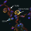
TLR2 and CD14 in the lungs of a rabbit. TLR2 is labeled red and CD14 is labeled green. Colocalization of TLR2 and CD14 is shown in yellow. TLR2 is visible on the alveolar epithelium and on alveolar macrophages in the airspace. CD14 is visible on alveolar macrophages, and neutrophils in the intravascular and alveolar space. The bright yellow alveolar macrophage shows high levels of expression of both TLR2 and CD14. Similar results are found when the sections are labeled for TLR4 and CD14.
<b>Figure 4.</b>
Figure 4.
(A–D) Clearance of gram-positive and gram-negative bacteria from the lungs and spleens of mice deficient in the MyD88 intracellular adaptor protein, which mediates intracellular signaling from TLRs. Mice deficient in MyD88 had markedly impaired clearance of Pseudomonas aeruginosa from the lungs, and died 24 h after aerosol exposure, but survived the challenge with the gram-positive organism, Staphylococcus aureus. CFU = colony-forming units. Reprinted by permission from Reference .
<b>Figure 5.</b>
Figure 5.
High- and low-response phenotypes in the human population using LPS as the defined stimulus in whole blood ex vivo. Peripheral venous blood from normal human volunteers was stimulated with LPS (10 ng/ml) for 6 h and cytokines were measured in the plasma supernatants. The results for each cytokine analysis were plotted in rank order and the dots for each subject were connected across all of the cytokines. Yellow lines denote consistently high responders, and blue lines denote consistently low responders. Other patterns are also visible in the background lines from the other subjects. IL = interleukin; TNF = tumor necrosis factor; MCP = monocyte chemotactic protein. Reprinted by permission from Reference .

References

    1. Beutler B. Innate immunity: an overview. Mol Immunol 2004;40:845–859. - PubMed
    1. Silverstein AM. History of immunology: cellular versus humoral immunity: determinants and consequences of an epic 19th century battle. Cell Immunol 1979;42:208–221. - PubMed
    1. Guo RF, Ward PA. Role of C5a in inflammatory responses. Annu Rev Immunol 2005;23:821–852. - PubMed
    1. Super M, Thiel S, Lu J, Levinsky RJ, Turner MW. Association of low levels of mannan-binding protein with a common defect of opsonisation. Lancet 1989;2:1236–1239. - PubMed
    1. Schumann RR, Leong SR, Flaggs GW, Gray PW, Wright SD, Mathison JC, Tobias PS, Ulevitch RJ. Structure and function of lipopolysaccharide binding protein. Science 1990;249:1429–1431. - PubMed

Publication types

Substances

LinkOut - more resources