. 2005 Nov;1(6):e66.
doi: 10.1371/journal.pcbi.0010066. Epub 2005 Nov 25.

Refining protein subcellular localization

Affiliations

Refining protein subcellular localization

Michelle S Scott et al. PLoS Comput Biol. 2005 Nov.

Abstract

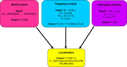
The study of protein subcellular localization is important to elucidate protein function. Even in well-studied organisms such as yeast, experimental methods have not been able to provide a full coverage of localization. The development of bioinformatic predictors of localization can bridge this gap. We have created a Bayesian network predictor called PSLT2 that considers diverse protein characteristics, including the combinatorial presence of InterPro motifs and protein interaction data. We compared the localization predictions of PSLT2 to high-throughput experimental localization datasets. Disagreements between these methods generally involve proteins that transit through or reside in the secretory pathway. We used our multi-compartmental predictions to refine the localization annotations of yeast proteins primarily by distinguishing between soluble lumenal proteins and soluble proteins peripherally associated with organelles. To our knowledge, this is the first tool to provide this functionality. We used these sub-compartmental predictions to characterize cellular processes on an organellar scale. The integration of diverse protein characteristics and protein interaction data in an appropriate setting can lead to high-quality detailed localization annotations for whole proteomes. This type of resource is instrumental in developing models of whole organelles that provide insight into the extent of interaction and communication between organelles and help define organellar functionality.


Conflict of interest statement

Competing interests. The authors have declared that no competing interests exist.

Figures

Figure 1
Figure 1. Structure of the PSLT2 Bayesian Network
The PSLT2 predictor is composed of three independent modules that can predict localization individually or in combination: the motif, targeting, and interaction modules. Each module can be characterized by the protein information used as input and the localization probabilities (for all compartments [C]) that are generated as output. The motif module accepts combinations of InterPro motifs (M) as input. The targeting module considers the presence of mitochondrial targeting signals (Mi), signal peptides/anchors (Si; S, signal peptide; A, signal anchor; Q, neither), GPI anchors (G), and the number of transmembrane domains (Tm) to predict localization. The interaction module considers the three compartments to which are localized the largest number of interactions partners (see Materials and Methods for more details). The full network (illustrated as the localization module) takes into account the output of all three modules to predict the probability of localization to all compartments (C).
Figure 2
Figure 2. Comparison between PSLT2, TRIPLES, and YeastGFP Datasets
Panels A through C represent an illustration of the Pearson correlation for the probability of localization between all compartment pairs for each pair of datasets (see Materials and Methods for details). SecPath, secretory pathway (ER and Golgi); Cyt, cytosol; Nuc, nucleus; Mit, mitochondrial or peroxisomal; PM & P, plasma membrane and periphery (including secreted and vacuolar proteins).
Figure 3
Figure 3. Sub-Compartmental Prediction Scheme
Proteins are predicted to be localized in one specific sub-compartment by first considering the most likely PSLT2 compartment (blue boxes). Further decisions depend on the PSLT2 second most likely compartment prediction (orange boxes) and targeting information (green boxes). Once all information has been analyzed, the protein is predicted to be in one of 18 sub-compartments (pink boxes). When the second most likely sub-compartments are considered, the default prediction is shown with a star (this branch of the tree is used in particular when proteins have no second most likely compartment as predicted by PSLT2). Pero, peroxisome; Vac, vacuole; Cyt, cytosolic; memb, membrane; TMD, number of transmembrane domains in protein; ER, endoplasmic reticulum; GPI, presence of GPI anchor; Nuc, nuclear; PM, plasma membrane; Mito, mitochondria; S, signal peptide; A, signal anchor; Q, neither signal peptide nor signal anchor.
Figure 4
Figure 4. Localizome–Interactomes
The protein–protein interaction maps for all proteins in the secretory pathway (B) or all proteins in the ER (C). Proteins are depicted as circles coloured according to their predicted sub-compartmental localization, as specified in the legend in (A). Interactions are shown as lines between proteins.

References

    1. Kumar A, Agarwal S, Heyman JA, Matson S, Heidtman M, et al. Subcellular localization of the yeast proteome. Genes Dev. 2002;16:707–719. - PMC - PubMed
    1. Huh WK, Falvo JV, Gerke LC, Carroll AS, Howson RW, et al. Global analysis of protein localization in budding yeast. Nature. 2003;425:686–691. - PubMed
    1. Wiemann S, Arlt D, Huber W, Wellenreuther R, Schleeger S, et al. From ORFeome to biology: A functional genomics pipeline. Genome Res. 2004;14:2136–2144. - PMC - PubMed
    1. Davis TN. Protein localization in proteomics. Curr Opin Chem Biol. 2004;8:49–53. - PubMed
    1. Scott MS, Thomas DY, Hallett MT. Predicting subcellular localization via protein motif co-occurrence. Genome Res. 2004;14:1957–1966. - PMC - PubMed

Publication types

MeSH terms

LinkOut - more resources