

Review
doi: 10.1172/JCI26987.
Thrombus formation in vivo
Affiliations
- PMID: 16322780
- PMCID: PMC1297262
- DOI: 10.1172/JCI26987
Item in Clipboard
Review
Thrombus formation in vivo
J Clin Invest.
2005 Dec.
Abstract
To examine thrombus formation in a living mouse, new technologies involving intravital videomicroscopy have been applied to the analysis of vascular windows to directly visualize arterioles and venules. After vessel wall injury in the microcirculation, thrombus development can be imaged in real time. These systems have been used to explore the role of platelets, blood coagulation proteins, endothelium, and the vessel wall during thrombus formation. The study of biochemistry and cell biology in a living animal offers new understanding of physiology and pathology in complex biologic systems.
Figures
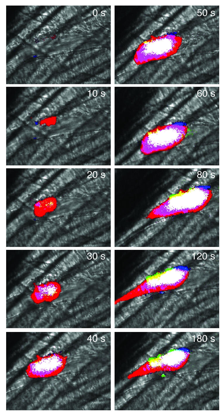
Birth of a thrombus. Intravital wide-field imaging of platelet, tissue factor, and fibrin deposition in the developing thrombus of a living WT mouse following endothelial injury. Blood flow is from right to left. Platelets, tissue factor, and fibrin were labeled using fluorescently tagged antibodies directed at CD41, tissue factor, and human fibrin, respectively. These components were imaged in 3 separate fluorescence channels. A black and white brightfield image indicates the histologic context of the composite image. To simplify analysis of the composite image, the dynamic range of the intensity of each pseudocolor was minimized. Red, platelets; green, tissue factor; blue, fibrin; yellow, platelets plus tissue factor; turquoise, tissue factor plus fibrin; magenta, platelets plus fibrin; white, platelets plus fibrin plus tissue factor.
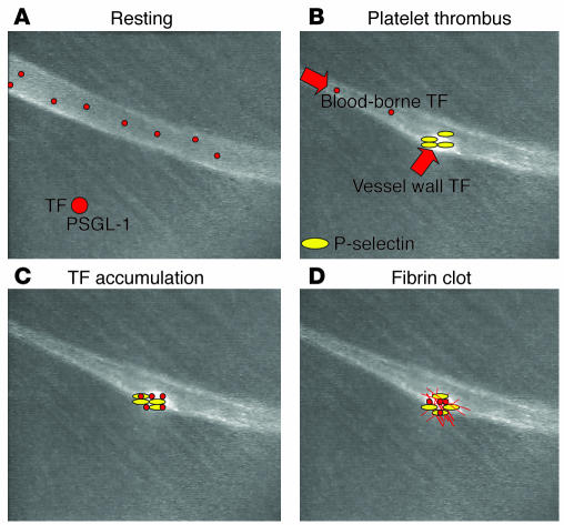
Model of P-selectin/PSGL-1–mediated tissue factor accumulation during thrombus formation. (A) Leukocyte microparticles (red) circulate constitutively in the blood under resting conditions. These microparticles express tissue factor (TF) and PSGL-1 on their surface. (B) Vessel wall tissue factor is expressed in response to laser-induced injury, leading to platelet activation and the subsequent expression of P-selectin on the stimulated platelets incorporated into the developing thrombus (initiation phase). (C) Blood-borne tissue factor associated with microparticles accumulates on the platelet thrombus through the binding of platelet P-selectin and microparticle PSGL-1. (D) Concentration of blood-borne tissue factor into the thrombus initiates thrombin generation and fibrin clot propagation within the thrombus (propagation phase).
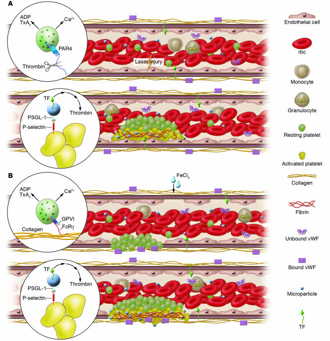
Experimental models of thrombosis. Platelets, red blood cells, monocytes, and granulocytes circulate in blood whereas endothelial cells line the vessel wall. Plasma proteins, including vWF, fibrinogen and other coagulation proteins, and microparticles are also present in the circulation. (A) Upon laser-induced injury of the vessel wall, vWF mediates the interaction of platelets with the endothelium. Tissue factor in the vessel wall leads to thrombin generation. Thrombin activates mouse platelets via the PAR4 receptor (inset). Activated platelets undergo calcium mobilization and the release of ADP and thromboxane A2 (TxA2) to accelerate platelet recruitment and activation and the formation of a platelet thrombus. These platelets express P-selectin, and leukocyte microparticles expressing PSGL-1 and tissue factor accumulate in the thrombus through the interaction of P-selectin with PSGL-1 (inset). The concentration of tissue factor initiates coagulation, the generation of more thrombin, and the propagation of a fibrin clot. (B) Upon vessel wall oxidative injury with ferric chloride, the endothelium is denuded and the subendothelial matrix exposed. Platelets interact with the matrix via GPIb-V-IX and αIIbβ3 on the platelet membrane and collagen and vWF in the matrix. Glycoprotein VI (GPVI) binding to collagen is required for platelet activation, and activated platelets undergo calcium mobilization and the release of ADP and thromboxane A2 (inset) to accelerate platelet recruitment and activation and the formation of a thrombus. These platelets express P-selectin, and microparticles expressing PSGL-1 and tissue factor accumulate in the thrombus through the interaction of P-selectin with PSGL-1 (inset). The concentration of tissue factor leads to coagulation, the generation of more thrombin, and the propagation of a fibrin clot.
References
-
- Furie B, Furie BC. The molecular basis of blood coagulation. Cell. 1988;53:505–518. - PubMed
-
- Ruggeri ZM. Platelets in atherothrombosis. Nat. Med. 2002;8:1227–1234. - PubMed
-
- Goto S. Understanding the mechanism of platelet thrombus formation under blood flow conditions and the effect of new antiplatelet agents. Curr. Vasc. Pharmacol. 2004;2:23–32. - PubMed
-
- Andrews RK, Berndt MC. Platelet physiology and thrombosis. Thromb. Res. 2004;114:447–453. - PubMed
Publication types
MeSH terms
Substances
LinkOut - more resources
Full Text Sources
Other Literature Sources
Medical
