Comparative Study
. 2005 Dec;242(6):859-67, discussion 867-8.
doi: 10.1097/01.sla.0000189572.02554.2c.

In vitro osteogenic differentiation and in vivo bone-forming capacity of human isogenic jaw periosteal cells and bone marrow stromal cells

Affiliations
Comparative Study

In vitro osteogenic differentiation and in vivo bone-forming capacity of human isogenic jaw periosteal cells and bone marrow stromal cells

Claude Jaquiéry et al. Ann Surg. 2005 Dec.

Abstract

Objective: To compare the in vitro osteogenic differentiation and in vivo ectopic bone forming capacity of human bone marrow stromal cells (BMSCs) and jaw periosteal cells (JPCs), and to identify molecular predictors of their osteogenic capacity.

Summary background data: JPC could be an appealing alternative to BMSC for the engineering of cell-based osteoinductive grafts because of the relatively easy access to tissue with minimal morbidity. However, the extent of osteogenic capacity of JPC has not yet been established or compared with that of BMSC.

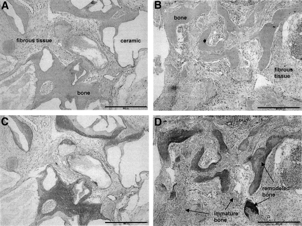
Methods: BMSCs and JPCs from the same donors (N = 9), expanded for 2 passages, were cultured for 3 weeks in osteogenic medium either in monolayers (Model I) or within 3-dimensional porous ceramic scaffolds, following embedding in fibrin gel (Model II). Cell-fibrin-ceramic constructs were also implanted ectopically in nude mice for 8 weeks (Model III). Cell differentiation in vitro was assessed biochemically and by real-time RT-PCR. Bone formation in vivo was quantified by computerized histomorphometry.

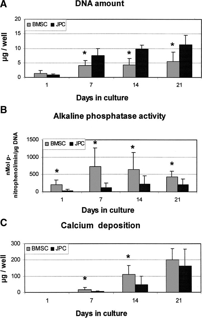
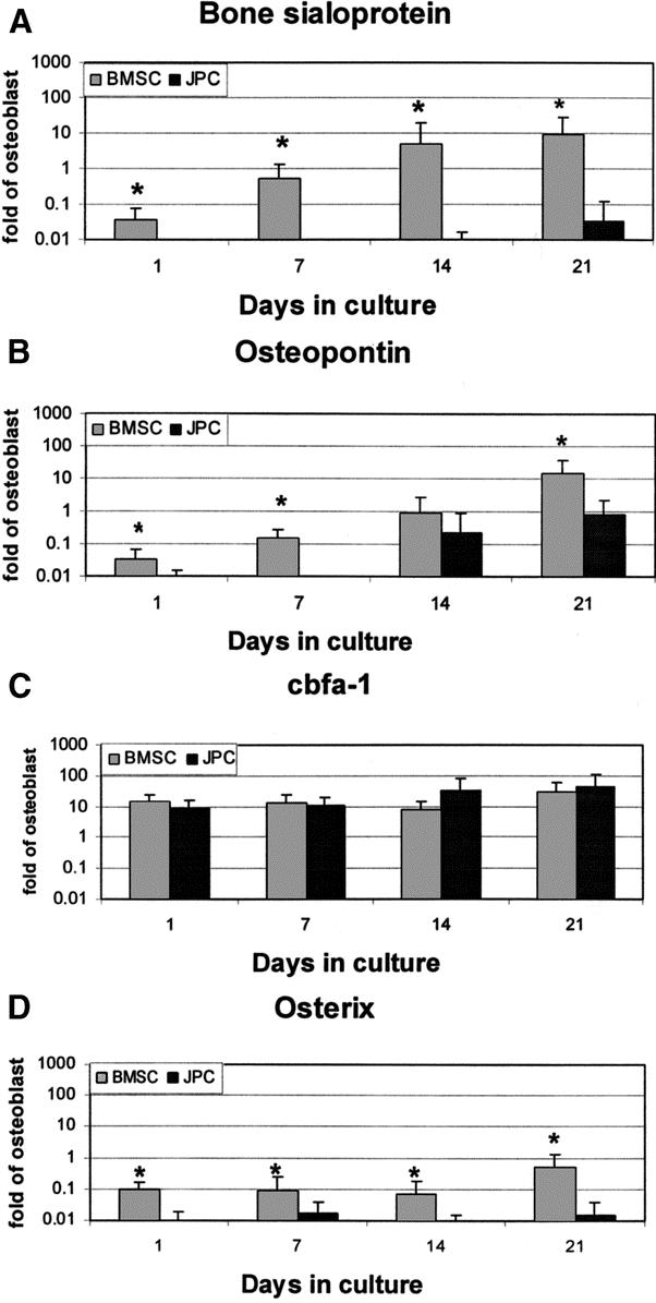
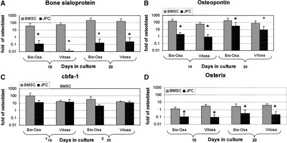
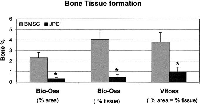
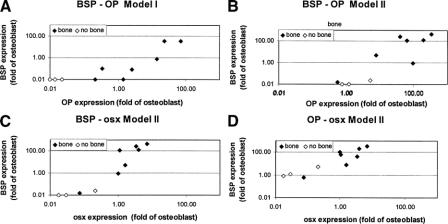
Results: JPCs had lower alkaline phosphatase activity, deposited smaller amounts of calcium (Model I), and expressed lower mRNA levels of bone sialoprotein, osteopontin, and osterix (Models I and II) than BMSCs. JPCs produced ectopic bone tissue at lower frequency and amounts (Model III) than BMSCs. Bone sialoprotein, osteopontin, and osterix mRNA levels by BMSCs or JPCs in Model II were markedly higher than in Model I and significantly more expressed by cells that generated bone tissue in Model III.

Conclusions: Our data indicate that JPCs, although displaying features of osteogenic cells, would not be as reliable as BMSCs for cell-based bone tissue engineering, and suggest that expression of osteoblast-related markers in vitro could be used to predict whether cells would be osteoinductive in vivo.


Figures

None
FIGURE 1. Biochemical analyses during cell differentiation in Model I. Human bone marrow stromal cells (BMSC) and jaw periosteal cells (JPC) were cultured in Model I (2D cultures) and assessed at different time points for the amount of DNA (A), alkaline phosphatase activity (B), and deposition of calcium (C). *Statistically significant difference (P < 0.01) between BMSC and JPC cultures.
None
FIGURE 2. Expression of osteoblast-related genes in Model I. Human bone marrow stromal cells (BMSC) and jaw periosteal cells (JPC) were cultured in Model I (2D cultures) and assessed at different time points for the mRNA expression of bone sialoprotein-I (A), osteopontin (B), cbfa-1 (C), and osterix (D). Values, normalized to 18 s, were expressed as fold difference from those previously measured in human osteoblasts. *Statistically significant differences (P < 0.01) between BMSC and JPC cultures.
None
FIGURE 3. Expression of osteoblast-related genes in Model II. Human bone marrow stromal cells (BMSC) and jaw periosteal cells (JPC) were cultured in Model II (3D cultures) using Bio-Oss or Vitoss scaffolds and assessed at 10 or 20 days for the mRNA expression of bone sialoprotein-I (A), osteopontin (B), cbfa-1 (C), and osterix (D). Values, normalized to 18 s, were expressed as fold difference from those previously measured in human osteoblasts. *Statistically significant differences (P < 0.01) between BMSC and JPC cultures.
None
FIGURE 4. Histology of bone tissue formation in Model III. Representative cross sections of constructs generated in Model III by bone marrow stromal cells using Bio-Oss (A, C) or Vitoss (B, D) scaffolds, stained by hematoxylin/eosin (A, B) or Masson/Trichrom (C, D). Empty spaces correspond to decalcified Bio-Oss. Vitoss was almost completely resorbed at this stage. In sections stained by Masson/Trichrome, a red color indicates remodeled and lamellar bone, whereas a blue color indicates immature and freshly deposited bone. Bone formation by jaw periosteal cells displayed similar histologic patterns. Bar = 0.5 mm.
None
FIGURE 5. Quantification of bone tissue formation in Model III. Bone tissue formation by human bone marrow stromal cells (BMSC) or jaw periosteal cells (JPC) generated in Model III using Bio-Oss or Vitoss was quantified by computerized histomorphometry. Bone tissue amount is expressed as a percentage of the total implant area (% area) or of the total space available for tissue ingrowth (% tissue). The 2 percentages were equivalent when Vitoss was used because of the almost complete resorption of the material. *Statistically significant differences (P < 0.01) between BMSC and JPC constructs.
None
FIGURE 6. Correlation between the expression of osteoblast-related genes. Graphical representation of the statistically significant correlations (Table 1) between the mRNA expression levels of bone sialoprotein-I (BSP), osteopontin (OP), and osterix (osx) by bone marrow stromal cells or jaw periosteal cells from each donor. Correlation between BSP-OP in Model I (A) and Model II (B), or between BSP-osx (C) and OP-osx (D) in Model II. The graphs also indicate whether single cell cultures formed or not bone tissue in Model III.

References

    1. Schnurer SM, Gopp U, Kuhn KD, et al. Bone substitutes. Orthopade. 2003;32:2–10. - PubMed
    1. Jaquiery C, Rohner D, Kunz C, et al. Reconstruction of maxillary and mandibular defects using prefabricated microvascular fibular grafts and osseointegrated dental implants: a prospective study. Clin Oral Implants Res. 2004;15:598–606. - PubMed
    1. Hartman EH, Spauwen PH, Jansen JA. Donor-site complications in vascularized bone flap surgery. J Invest Surg. 2002;15:185–197. - PubMed
    1. Bianco P, Gehron RP. Marrow stromal stem cells. J Clin Invest. 2000;105:1663–1668. - PMC - PubMed
    1. Owen M. Marrow stromal stem cells. J Cell Sci Suppl. 1988;10:63–76. - PubMed

Publication types

MeSH terms