Comparative Study
. 2005 Dec 13;102(50):18147-52.
doi: 10.1073/pnas.0509073102. Epub 2005 Dec 5.

The genome of Salinibacter ruber: convergence and gene exchange among hyperhalophilic bacteria and archaea

Affiliations
Comparative Study

The genome of Salinibacter ruber: convergence and gene exchange among hyperhalophilic bacteria and archaea

E F Mongodin et al. Proc Natl Acad Sci U S A. .

Abstract

Saturated thalassic brines are among the most physically demanding habitats on Earth: few microbes survive in them. Salinibacter ruber is among these organisms and has been found repeatedly in significant numbers in climax saltern crystallizer communities. The phenotype of this bacterium is remarkably similar to that of the hyperhalophilic Archaea (Haloarchaea). The genome sequence suggests that this resemblance has arisen through convergence at the physiological level (different genes producing similar overall phenotype) and the molecular level (independent mutations yielding similar sequences or structures). Several genes and gene clusters also derive by lateral transfer from (or may have been laterally transferred to) haloarchaea. S. ruber encodes four rhodopsins. One resembles bacterial proteorhodopsins and three are of the haloarchaeal type, previously uncharacterized in a bacterial genome. The impact of these modular adaptive elements on the cell biology and ecology of S. ruber is substantial, affecting salt adaptation, bioenergetics, and photobiology.


Figures

Fig. 1.
Fig. 1.
Normalized distribution of pI values at 0.2 intervals for predicted ORFs in Haloarcula marismortui (purple), Halobacterium sp. NRC-1 (red), Salinibacter (blue), C. tepidum (green), and B. fragilis (cyan). Predicted pI values of the proteins were calculated by using NeuroGadgets Bioinformatics Web Service.
Fig. 2.
Fig. 2.
The nature and number of potential lateral gene transfers involving Salinibacter. Black bars correspond to the number of Salinibacter genes for which this genome is the best blast hit. Gray bars correspond to the number of genes for which maximum likelihood phylogenetic analyses (18) indicated a sister relationship with this genome and, thus, a potential LGT between the two species. The pie charts show whether LGT between Salinibacter and the indicated genome was supported and, if so, its direction. Of those trees, the blue fraction support transfer from the indicated genome to Salinibacter, the chartreuse fraction support transfer from Salinibacter to this genome, the green fraction support transfer between Salinibacter and this genome for which no decision on direction can be made, and the red fraction do not support transfer between Salinibacter and this genome. When several genomes made up the sister group to or from which LGT might be inferred, no simple LGT event was assumed. Thus, complex or ancient events were not scored here.
Fig. 3.
Fig. 3.
A schematic representation of the hypersalinity island identified in the genome of Salinibacter. Genes are color-coded with respect to their closest matches in blast sequence similarity searches: haloarchaea, red; cyanobacteria, green; methanogenic archaea, yellow; firmicutes, blue.
Fig. 4.
Fig. 4.
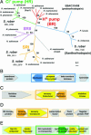
The phylogeny and genomic context of Salinibacter rhodopsin genes. (A) Maximum likelihood phylogeny of rhodopsin genes inferred from 212 conserved amino acid positions by phyml (18). The evolutionary model and parameters used in the analyses were WAG + F + γ + I. Support for nodes correspond to 100 bootstrap replicates. Scale bar indicates the number of positions per site. (B-E) Gene context of Salinibacter rhodopsin genes. Darker boxes suggest possible cotranscribed and/or coregulated gene sets (same transcription direction and <250 bp separating any two adjacent genes within the box) Genes colored in yellow indicate those with homologs in halophilic archaea with blast expect value <1-10.

References

    1. Anton, J., Rossello-Mora, R., Rodriguez-Valera, F. & Amann, R. (2000) Appl. Environ. Microbiol. 66, 3052-3057. - PMC - PubMed
    1. Anton, J., Oren, A., Benlloch, S., Rodriguez-Valera, F., Amann, R. & Rossello-Mora, R. (2002) Int. J. Syst. Evol. Microbiol. 52, 485-491. - PubMed
    1. Oren, A., Heldal, M., Norland, S. & Galinski, E. A. (2002) Extremophiles 6, 491-498. - PubMed
    1. Oren, A. & Mana, L. (2002) Extremophiles 6, 217-223. - PubMed
    1. Sher, J., Elevi, R., Mana, L. & Oren, A. (2004) FEMS Microbiol. Lett. 232, 211-215. - PubMed

Publication types

Substances