Comparative Study
. 2005 Dec 13;102(50):18183-8.
doi: 10.1073/pnas.0506535102. Epub 2005 Dec 5.

Expression pattern of the transcription factor Olig2 in response to brain injuries: implications for neuronal repair

Affiliations
Comparative Study

Expression pattern of the transcription factor Olig2 in response to brain injuries: implications for neuronal repair

Annalisa Buffo et al. Proc Natl Acad Sci U S A. .

Abstract

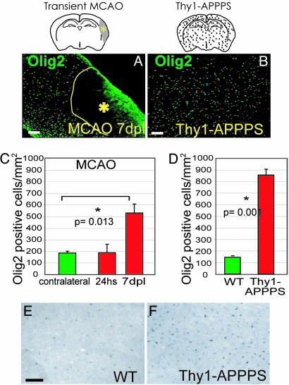
Despite the presence of neural stem cells and ongoing neurogenesis in some regions of the adult mammalian brain, neurons are not replaced in most brain regions after injury. With the aim to unravel factors contributing to the failure of neurogenesis in the injured cerebral cortex, we examined the expression of cell fate determinants after acute brain injuries, such as stab wound or focal ischemia, and in a model of chronic amyloid deposition. Although none of the neurogenic factors, such as Pax6, Mash1, Ngn2, was detected in the injured parenchyma, we observed a strong up-regulation of the bHLH transcription factor Olig2, but not Olig1, upon acute and chronic injury. To examine the function of Olig2 in brain lesion, we injected retroviral vectors containing a dominant negative form of Olig2 into the lesioned cortex 2 days after a stab wound. Antagonizing Olig2 function resulted in a significant number of infected cells generating immature neurons that were not observed after injection of the control virus. These data, therefore, imply Olig2 as a repressor of neurogenesis in cells reacting to brain injury and open innovative perspectives toward evoking endogenous neuronal repair.


Figures

Fig. 1.
Fig. 1.
Increase in the number of Olig2-immunoreactive cells after a stab wound. (A and B) Frontal sections of the gray matter (GM) in the adult mouse neocortex stained for Olig2. Note the increase in the number of Olig2+ cells after a stab wound (s-w, B; yellow line indicates the lesion site, dashed contours outline some unspecific/autofluorescent staining, and arrowheads point to some Olig2+ cells) compared to the intact cortex (A, arrowheads indicate Olig2+ cells) as quantified in the histogram (C). *, statistically significant differences; hs, hours; dpl, days after lesion. In situ hybridization reveals Olig2 mRNA up-regulation after a stab wound (E) compared to the intact cortex (D). (Scale bars: 100 μm.)
Fig. 2.
Fig. 2.
Identification of Olig2-immunoreactive cells after stab-wound and proliferation analysis. (AE) Frontal sections of the intact neocortical GM immunostained as indicated. Olig2 immunoractivity was detected in CC1+ oligodendrocytes (A), NG2+ glial precursors (B), and S100β+ astrocytes (C). Arrows point to colabeled cells, and arrowheads point to cells devoid of Olig2. (D) Olig2 and Sox10 coexpression (arrows) in the cortical GM are shown. (E) The micrograph depicts Olig2+ nuclei triple labeled for BrdUrd (red) and a mixture of antibodies (in blue: NG2, CC1, and S100β) close to the stab-wound track: Several proliferating Brdu+/Olig2+ cells are lineage marker-negative (arrow). Histograms in F depict the proportions of cell types in the Olig2+ population in the intact cortex and after a stab wound, whereas in G, the percentage of Olig2+ cells amongst different glial populations is shown. The fractions of Olig2+ astrocytes increased at 7 dpl (P = 0.012) compared to the intact situation. The opposite was observed for the NG2+ cells in comparison with the intact cortex (P = 0.003 at 3 days; P = 0.042 at 7 days) and the proportion of Olig2+/CC1+ oligodendrocytes rose 7 dpl (P = 0.044). (H) Olig2/BrdUrd colabeled cells (indicated also as percentages) are shown over the density of BrdUrd-positive cells in the intact and stab-lesioned cortex (2-h BrdUrd pulse). (I) The composition of BrdUrd-positive cells is depicted after 2 h (Left) or several days of BrdUrd pulse (Right). (Scale bars: AC and E, 20 μm; D, 100 μm.)
Fig. 3.
Fig. 3.
Repression of Olig2 function after a stab wound. (A) The viral injection experimental design is illustrated: Animals were injected with either the GFP or the Olig2VP16 virus 2 days after a stab wound and killed later. The micrograph (B) illustrates the distribution of GFP+-infected cells in the cortex 7 days after control virus injection (GM, gray matter; WM, white matter). (CI) GFP+ cells 7 days after viral transduction (as specified in the images) stained for the antigens indicated. (F) Arrow points to a marker-negative infected cell. The micrographs (II′′′) show examples of Pax6 and dcx coexpression in Olig2VP16-infected cells. The graphs in JL illustrate the phenotypes of virus-infected cells as indicated (three mice analyzed for cell type identification at each time point; 100–200 analyzed cells unless differently indicated in the text; dpi, days after injection). (Scale bars: B, 200 μm; CG,20 μm; H,40 μm; II′′′,10 μm.)
Fig. 4.
Fig. 4.
Increase in the number of Olig2-immunoreactive cells after focal ischemia (MCAO) and in the Thy1-APPPS cortex. (A and B) Anti-Olig2 immunostaining of frontal sections of the cortical GM shows an increased number of Olig2+ cells after MCAO (A) and after amyloid plaque formation (B) compared to the control cortex (see Fig. 1 A). Histograms in C and D display the corresponding quantifications. hs, hours; dpl, days after lesion. In situ hybridization reveals Olig2 mRNA up-regulation in the Thy1-APPPS (F) compared to the wild-type cortex (E). (Scale bars: 100 μm.)

References

    1. Doetsch, F., Caille, I., Lim, D. A., Garcia-Verdugo, J. M. & Alvarez-Buylla, A. (1999) Cell 97, 703-716. - PubMed
    1. Garcia, A. D., Doan, N. B., Imura, T., Bush, T. G. & Sofroniew, M. V. (2004) Nat. Neurosci. 7, 1233-1241. - PubMed
    1. Fawcett, J. W. & Asher, R. A. (1999) Brain Res. Bull. 49, 377-391. - PubMed
    1. Ridet, J. L., Malhotra, S. K., Privat, A. & Gage, F. H. (1997) Trends Neurosci. 20, 570-577. - PubMed
    1. Hartfuss, E., Galli, R., Heins, N. & Götz, M. (2001) Dev. Biol. 229, 15-30. - PubMed

Publication types

Substances