. 2006 May 11;25(20):2943-9.
doi: 10.1038/sj.onc.1209310.

Cyclin-dependent kinase 2 is dispensable for normal centrosome duplication but required for oncogene-induced centrosome overduplication

Affiliations

Cyclin-dependent kinase 2 is dispensable for normal centrosome duplication but required for oncogene-induced centrosome overduplication

A Duensing et al. Oncogene. .

Abstract

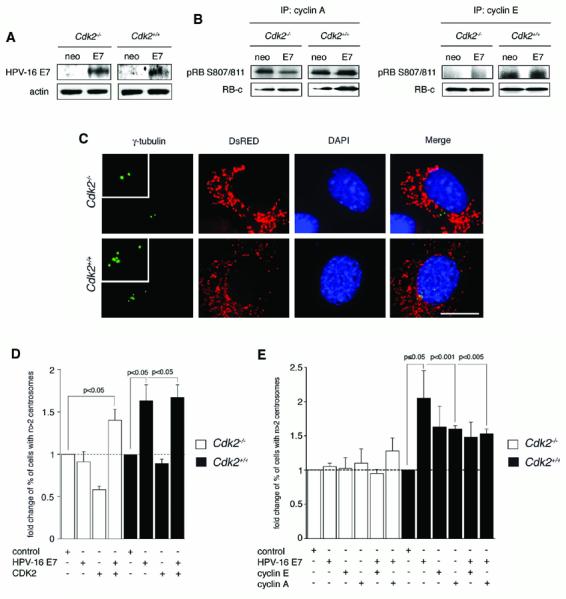
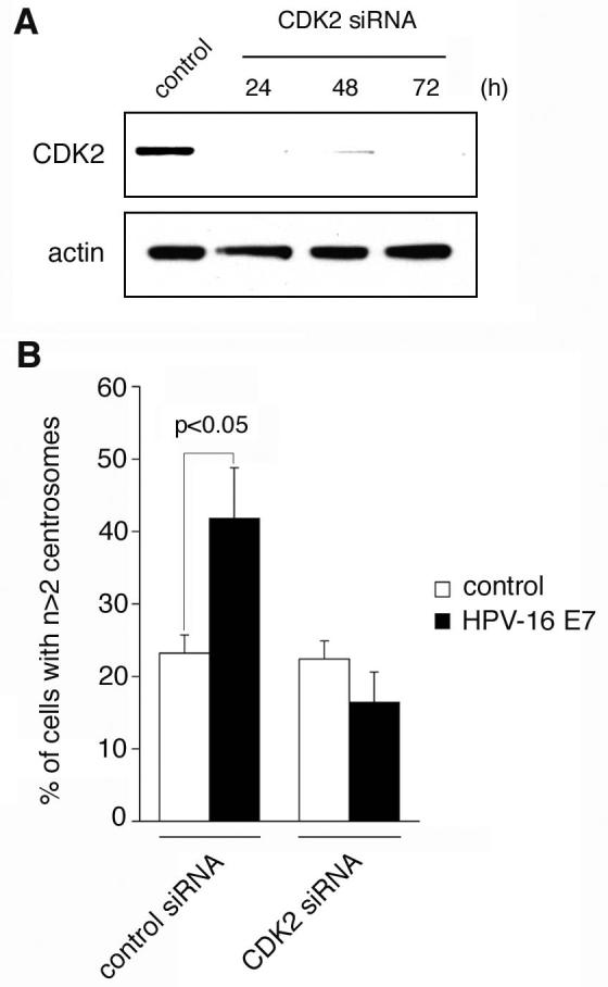
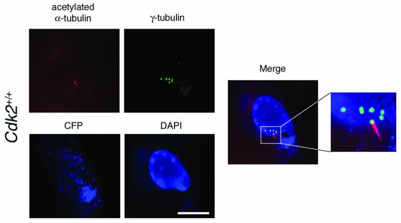
Cyclin-dependent kinase 2 (CDK2) has been proposed to function as a master regulator of centrosome duplication. Using mouse embryonic fibroblasts (MEFs) in which Cdk2 has been genetically deleted, we show here that CDK2 is not required for normal centrosome duplication, maturation and bipolar mitotic spindle formation. In contrast, Cdk2 deficiency completely abrogates aberrant centrosome duplication induced by a viral oncogene. Mechanistically, centrosome overduplication in MEFs wild-type for Cdk2 involves the formation of supernumerary immature centrosomes. These results indicate that normal and abnormal centrosome duplication have significantly different requirements for CDK2 activity and point to a role of CDK2 in licensing centrosomes for aberrant duplication. Furthermore, our findings suggest that CDK2 may be a suitable therapeutic target to inhibit centrosome-mediated chromosomal instability in tumor cells.


Figures

Figure 1
Figure 1. CDK2-independent centrosome duplication and maturation in Cdk2−/− MEFs
(A) Cdk2−/− MEFs were analyzed by immunofluorescence microscopy for γ-tubulin, a commonly used centrosome marker, to detect centrosomes (arrowheads) during interphase (top panels) and mitosis (bottom panels). Cells plated on coverslips were washed briefly in phosphate-buffered saline (PBS) and then fixed in 4% paraformaldehyde/PBS (pH 7.0) for 10 min at room temperature. Cells were permeabilized with 1% Triton-X 100 (Sigma) in PBS for 10 min at room temperature and non-specific staining was blocked with 10% normal donkey serum (Jackson Immunoresearch) in distilled water for 15 min at room temperature. Cells were then incubated with a primary mouse monoclonal anti-γ-tubulin antibody (Sigma) at a 1:1000 dilution in PBS overnight at 4°C followed by a FITC-conjugated donkey anti-mouse secondary antibody (Jackson Immunoresearch) at a 1:100 dilution in PBS for 2 h at 37°C. Cells were mounted with DAPI (Vector Laboratories) and analyzed using an Olympus AX70 epifluorescence microscope equipped with a SpotRT digital camera. Pictures were transferred to Adobe Photoshop for print-out. Scale bar equals 10 μm. (B) Quantification of centrosome numbers following γ-tubulin staining in asynchronously growing Cdk2+/+ and Cdk2−/− MEFs. Each bar represents mean + standard error of three independent experiments and counts of at least one-hundred cells per experiment. (C) Immunofluorescence microscopy for acetylated α-tubulin and γ-tubulin reveals the primary cilium-carrying centrosome in a Cdk2−/− MEF after serum-deprivation for 24 h. For detection of primary cilia, cells were first stained for γ-tubulin using a rabbit primary antibody (Santa Cruz) at a 1:100 dilution in PBS followed by a FITC-labeled donkey anti-rabbit secondary antibody (Jackson Immunoresearch) at a 1:200 dilution in PBS and then incubated with an anti-acetylated α-tubulin antibody (Zymed) at a 1:100 dilution in PBS followed by a Rhodamine Red-conjugated donkey anti-mouse secondary antibody (Jackson Immunoresearch) at a 1:500 in PBS. Cells were then processed as described above. Scale bar equals 10 μm. (D) Quantification of primary cilium-carrying centrosomes in serum-starved Cdk2−/− and Cdk2+/+ MEFs in correlation to the total centrosome number per cell. Results from a representative experiment in which a total of n=186 cells were evaluated are shown.
Figure 2
Figure 2. CDK2 is required for centrosome overduplication induced by a viral oncogene
(A) Immunoblot analysis of HPV-16 E7 expression in Cdk2−/− and Cdk2+/+ MEFs compared to empty vector (neo) after transient transfection. Cells were transfected with 2 μg of plasmid DNA using Fugene 6 (Invitrogen) and processed for immunoblot analysis after 48 h. Whole cell lysates were prepared by scraping monolayer cells into ice-cold lysis buffer (1% NP-40, 50 mM Tris-Hcl pH 8.0, 100 mM sodium fluoride, 30 mM sodium pyrophosphate, 2 mM sodium molybdate, 5 mM EDTA, 2 mM sodium orthovanadate) containing 10 μg/ml aprotinin, 10 μg/ml leupeptin, 1 mM phenylmethylsulfonyl fluoride and additional sodium orthovanadate (2 mM). Lysates were incubated for 1 h with shaking at 4°C and then cleared by centrifugation for 30 min at 14,000 rpm at 4°C. SDS-gel electrophoresis and immunoblotting were performed using 4-12% Bis-Tris gels (Invitrogen) and a MiniTrans Blot Cell (Biorad Laboratories). Blot immunostains were detected by enhanced chemiluminescence (ECL, Amersham), exposed to X-ray film and developed in a Kodak X-OMAT 2000 film processor (Eastman Kodak). The pCDNA3-HPV-16 E7-HA construct was kindly provided by K. Munger (Harvard Medical School, Boston), empty pCDNA3-HA was kindly provided by Yuan Chang and Patrick Moore (University of Pittsburgh Cancer Institute, Pittsburgh). (B) In vitro activity of cyclin A- (left panels) or cyclin E- (right panels) associated kinases following transient transfection of Cdk2−/− and Cdk2+/+ MEFs with HPV-16 E7 or empty vector (neo). Kinase reactions were performed with 375 μg of protein. Immunoprecipitations using agarose-conjugated cyclin A polyclonal antibodies (H-432, Santa Cruz) or cyclin E polyclonal antibodies (M2, Santa Cruz) were performed overnight at 4°C. Immunocomplexes were washed three times in lysis buffer (1% NP-40, 50 mM Tris-Hcl pH 8.0, 100 mM sodium fluoride, 30 mM sodium pyrophosphate, 2 mM sodium molybdate, 5 mM EDTA, 2 mM sodium orthovanadate) and three times in kinase buffer (10 mM MgCl2, 25 mM Tris HCl, 100 μM ATP, at pH 7.4). Samples were then resuspended in 20 μl of kinase buffer containing 0.5 μg of recombinant C-terminal fragment of the retinoblastoma protein (RB-c) comprising amino acids 773-928 (Upstate). Phosphorylation of RB-c was determined using a phospho-specific antibody against serine residues 807/811 (Cell Signaling). Total RB-c was measured using an antibody recognizing the C-terminus of pRB (4H1, Cell Signaling). (C) Immunofluorescence analysis of Cdk−/− and Cdk2+/+ MEFs transiently transfected with HPV-16 E7. Cells were co-transfected with a plasmid encoding DsRED protein targeted to mitochondria (Clontech) at a 1:10 ratio to label transfected cells. Centrosomes were visualized by immunofluorescence staining for γ-tubulin using a mouse monoclonal antibody (see above). Nuclei were stained with DAPI. Scale bar equals 10 μm. (D) Quantification of fold changes of the percentage of Cdk2−/− or Cdk2+/+ MEFs with abnormal centrosome numbers (i.e., more than two per cell) after transient transfection with HPV-16 E7 or CDK2 either alone or in combination. For cotransfection experiments, 1 μg of each plasmid DNA was used. The CDK2 expression construct was kindly provided by P. Hinds (Tufts University, Boston). Results were normalized to the mean percentage of cells with abnormal centrosome numbers in empty vector-transfected MEFs (control). Each bar indicates mean of fold changes and standard error obtained from at least three independent experiments and assessment of at least fifty DsRED-positive cells per experiment. (D) Quantification of fold changes of the percentage of Cdk2−/− or Cdk2+/+ MEFs with abnormal centrosome numbers (i.e., more than two per cell) after transient transfection with cyclin E or cyclin A either alone or in combination with HPV-16 E7. For co-transfection experiments, 1 μg of each plasmid DNA was used. The cyclin E and cyclin A constructs were kindly provided by P. Hinds (Tufts University, Boston) Results were normalized to the mean percentage of cells with abnormal centrosome numbers in empty vector-transfected MEFs (control). Each bar indicates mean of fold changes and standard error obtained from at least three independent experiments and assessment of at least twenty-five DsRED-positive cells per experiment.
Figure 3
Figure 3. Small-interfering RNA (siRNA)-mediated knock-down of CDK2 prevents HPV-16 E7-induced abnormal centrosome duplication.
(A) Immunoblot analysis of wild-type MEFs for CDK2 expression following transfection with siRNA duplexes targeting CDK2 or control siRNAs. Two synthetic oligonucleotides targeting mouse CDK2 were used (5'-GGUGUACCCAGUACUGCCA-3' and 5'-GGAACUUAAUCACCCUAAU-3'; Silencer™, Ambion). Pre-designed control oligonucleotides were used as negative control (Silencer™ Negative, Ambion). Wild-type MEFs grown on coverslips in 60 mm plates with 2 ml DMEM free of antibiotics were transfected with 5 μl of 20 μM annealed duplexes using nucleofection (Amaxa). Whole cell lysates were prepared at the indicated time points and analyzed by immunoblotting as described above. Immunoblot for actin is shown to demonstrate loading of equal amounts of protein. (B) Quantification of the percentage of MEFs with abnormal centrosome numbers following siRNA-directed knock-down of CDK2 protein or transfection with control duplexes and transient transfection with HPV-16 E7 or empty vector. Cells were transfected with siRNAs as described above and transfected with HPV-16 E7 or controls plasmids after 24 h. Cells were co-transfected with DsRED and processed for immunofluorescence microscopy for γ-tubulin after additional 48 h. Mean and standard errors of at least three independent experiments with at least 50 DsRED-positive cells counted per experiment are shown.
Figure 4
Figure 4. CDK2 mediates excessive generation of immature centrosomes induced by the HPV-16 E7 oncoprotein
Immunofluorescence microscopy analysis for γ-tubulin and acetylated α-tubulin (see above) demonstrates multiple immature centrosomes in the presence of a single primary cilium-carrying centrosome in a HPV-16 E7-transfected cell. Cdk2+/+ MEFs were transiently transfected with HPV-16 E7 using cyan fluorescent protein (CFP) targeted to mitochondria (Clontech) at a 1:10 ratio as transfection marker. Pictures were taken from a representative experiment in which a total of n=53 transfected Cdk2+/+ MEFs were evaluated. Nucleus stained with DAPI. Scale bar equals 10 μm.

Similar articles

Cited by

References

    1. Albrecht-Buehler G, Bushnell A. Exp Cell Res. 1980;126:427–37. - PubMed
    1. Alvarez-Salas LM, Cullinan AE, Siwkowski A, Hampel A, DiPaolo JA. Proc Natl Acad Sci U S A. 1998;95:1189–94. - PMC - PubMed
    1. Berthet C, Aleem E, Coppola V, Tessarollo L, Kaldis P. Curr Biol. 2003;13:1775–85. - PubMed
    1. Bornens M. Curr. Opin. Cell Biol. 2002;14:25–34. - PubMed
    1. Brinkley BR. Trends Cell Biol. 2001;11:18–21. - PubMed

Publication types

Substances