. 2006 Feb;147(3):249-59.
doi: 10.1038/sj.bjp.0706551.

Differential phosphorylation and dephosphorylation of beta2-adrenoceptor sites Ser262 and Ser355,356

Affiliations

Differential phosphorylation and dephosphorylation of beta2-adrenoceptor sites Ser262 and Ser355,356

Varsha Iyer et al. Br J Pharmacol. 2006 Feb.

Abstract

Activated beta2-adrenoceptors are rapidly desensitized by phosphorylation of Ser262 by protein kinase A (PKA) and of Ser355,356 by G-protein-coupled receptor kinase (GRK). We sought to determine whether the phosphorylation and subsequent dephosphorylation of these sites had similar kinetics and requirements for receptor endocytosis. The phosphorylation of the PKA and GRK sites were measured using antibodies that recognize phosphoserine 262 and phosphoserine 355,356. Endocytosis in stably transfected HEK293 cells was blocked by inducible expression of dominant-negative dynamin-1 K44A or by treatment with hypertonic sucrose. The phosphorylation of the GRK site Ser355,356 during a 10 microM isoprenaline treatment rapidly reached a steady state, and the extent of kinetics of phosphorylation were unaffected by dynamin-1 K44A expression, and minimally by hypertonic sucrose. In contrast, phosphorylation of the PKA site Ser262 during a 10 microM isoprenaline treatment peaked after 2 min and then rapidly declined, while inhibition of endocytosis enhanced and prolonged phosphorylation. Treatment with 300 pM isoprenaline, a concentration too low to provoke endocytosis, also resulted in prolonged PKA site phosphorylation. The dephosphorylation of these sites was measured after removal of agonist. Significant dephosphorylation of phosphoserines 262 and 355,356 was observed under conditions of very low endocytosis, however dephosphorylation of the GRK site was greater if antagonist was present after removal of agonist. The results indicate that the kinetics of beta2-adrenoceptor GRK and PKA site phosphorylation are distinct and differently affected by endocytosis, and that receptor dephosphorylation can occur either at the plasma membrane or in internal compartments.


Figures
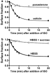
Figure 1
Figure 1
Inhibition of β2-adrenoceptor endocytosis. Cells were grown in 24-well clusters, then isoprenaline (10 μM) was added to triplicate wells for 1–20 min before the clusters were quickly chilled. The monolayers were washed and then incubated on ice with 6 nM [3H]CGP-12177 in DMEM-H for 60 min. The label was aspirated and cells were washed and lysed for scintillation counting. The fraction of receptors left on the surface (relative to no agonist) is plotted as a function of time. The curves were modeled as described in Morrison et al. (1996). (a) The 12β6 cells were treated with 0.5 M sucrose in HBSS or HBSS alone for 30 min prior to and during exposure to agonist (N=2). (b) EcR-βAR-dynKA cells were treated with 5 μM ponasterone or with vehicle for 72 h prior to the addition of agonist (N=9).
Figure 2
Figure 2
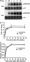
Phosphorylation of β2-adrenoceptors at the GRK site Ser355,356 after dominant-negative dynamin inhibition of endocytosis. EcR-βAR-DynKA cells were grown for 72 h in the presence of 5 μM ponasterone or vehicle alone. Isoprenaline was added to 10 μM for varying times, and then the cells were chilled and harvested in lysis buffer as described in the Methods. Equivalent protein was loaded in each lane for polyacrylamide gel electrophoresis (PAGE), then immunoblotted using the antibody against phosphoserine 355,356 (α-p355,356) and visualized by chemiluminescence. After stripping the blot, it was probed with antibody to the β2-adrenoceptor C-terminus (α-CT) to normalize phosphoreceptor levels to total receptors. (a) Representative blots; (b) data plotted as fold over basal and (c) data plotted as fraction of maximum phosphorylation (N=3).
Figure 3
Figure 3
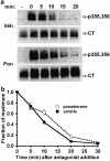
Dephosphorylation of β2-adrenoceptors at the GRK site Ser355,356 after dominant-negative dynamin inhibition of endocytosis. EcR-βAR-dynKA cells were grown for 72 h in the presence of 5 μM ponasterone or vehicle alone. Isoprenaline was added to 10 μM for 5 min, and then the cells were rapidly washed and the incubation continued at 37oC in fresh medium with 3 μM propranolol. At various times, the cells were chilled and harvested in lysis buffer as described in the Methods. Equivalent protein was loaded in each lane for PAGE, then immunoblotted using the antibody against phosphoserine 355,356 (α-p355,356) and visualized by chemiluminescence. After stripping the blot, it was probed with antibody to the β2-adrenoceptor C-terminus (α-CT) to normalize phosphoreceptor levels to total receptors. (a) Representative blots and (b) data plotted as fraction of maximum phosphorylation (N=3).
Figure 4
Figure 4
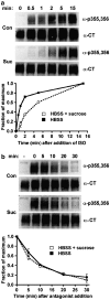
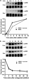
Phosphorylation and dephosphorylation of β2-adrenoceptors at the GRK site Ser355,356 after hypertonic sucrose treatment. The 12β6 cells were grown for 24 h, then treated with HBSS alone or HBSS+0.5 M sucrose at 37oC for 30 min. (a) Phosphorylation. Isoprenaline was added to 10 μM for varying times, and then the cells were chilled and harvested in lysis buffer as described in the Methods. (b) Dephosphorylation. After 5 min of 10 μM isoprenaline treatment, the cells were washed three times with medium, and then incubated for varying times in HBSS or HBSS+0.5 M sucrose containing 3 μM propranolol prior to harvest. For both (a) and (b), equivalent protein was loaded in each lane for PAGE, then immunoblotted using the antibody against phosphoserine 355,356 (α-p355,356) and visualized by chemiluminescence. After stripping the blot, it was probed with antibody to the β2-adrenoceptor C-terminus (α-CT) to normalize phosphoreceptor levels to total receptors. At the top of each panel are shown representative blots, with the data plotted as a fraction of maximum phosphorylation from three experiments shown in the lower panels (N=3).
Figure 5
Figure 5
Dephosphorylation of β2-adrenoceptors at the GRK site Ser355,356 after varying times of agonist exposure in the presence and absence of antagonist. (a) EcR-βAR-dynKA cells were grown for 72 h in the presence of 5 μM ponasterone or vehicle alone. Isoprenaline was added to 1 μM for either 5 min or 15 min, then the cells were washed extensively and incubated for 30 min in the presence or absence of 1 μM propranolol. The cells were chilled and harvested in lysis buffer, and Ser355,356 phosphorylation measured as described in the Methods. The levels of β2-adrenoceptor phosphorylation normalized to total receptor levels are shown as the percentage of maximum phosphorylation obtained after 5 or 15 min of agonist exposure. Values are the means±s.e.m. (N=3), where each experiment was performed in duplicate. *Significantly different (P<0.05, Student's two-tailed t-test) plus propranolol versus no propranolol. (b) HEK293 cells stably expressing HA-β2ARs (12β6 cells) were treated with HBSS or HBSS with 0.5 M sucrose for 30 min at 37oC, then with isoprenaline (1 μM) for either 5 or 15 min. The cells were washed extensively and incubated for 30 min in the presence or absence of 1 μM propranolol, then chilled and harvested in lysis buffer and Ser355,356 phosphorylation measured as described in the Methods. The levels of β2-adrenoceptor phosphorylation normalized to total receptor levels are shown as the percentage of maximum phosphorylation obtained after 5 or 15 min of agonist exposure. The error bars show the ranges of two determinations.
Figure 6
Figure 6
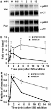
Phosphorylation of β2-adrenoceptors at the PKA site Ser262 after dominant-negative dynamin inhibition of endocytosis. EcR-βAR-dynKA cells were grown for 72 h in the presence of 5 μM ponasterone or vehicle alone. Isoprenaline was added to 10 μM for varying times, and then the cells were chilled and harvested in lysis buffer as described in the Methods. Equivalent protein was loaded in each lane for PAGE, then immunoblotted using the antibody against phosphoserine 262 (α-p262) and visualized by chemiluminescence. After stripping the blot, it was probed with antibody to the β2-adrenoceptor C-terminus (α-CT) to normalize phosphoreceptor levels to total receptors. (a) Representative blots (b) data plotted as fold over basal and (c) data plotted as fraction of maximum phosphorylation. *P<0.05 (Student's two-tailed t-test) (N=4).
Figure 7
Figure 7
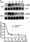
Dephosphorylation of β2-adrenoceptors at the PKA site Ser262 after dominant-negative dynamin inhibition of endocytosis. EcR-βAR-dynKA cells were grown for 72 h in the presence of 5 μM ponasterone or vehicle alone. Isoprenaline was added to 10 μM for 5 min, and then the cells were rapidly washed and the incubation continued at 37°C in fresh medium 3 μM propranolol. At various times, the cells were chilled and harvested in lysis buffer as described in the Methods. Equivalent protein was loaded in each lane for PAGE, then immunoblotted using the antibody against phosphoserine 262 (α-p262) and visualized by chemiluminescence. After stripping the blot, it was probed with antibody to the β2-adrenoceptor C-terminus (α-CT) to normalize phosphoreceptor levels to total receptors. (a) Representative blots and (b) data plotted as fraction of maximum phosphorylation (N=3).
Figure 8
Figure 8
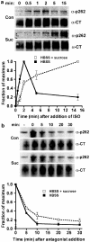
Phosphorylation and dephosphorylation of β2-adrenoceptors at the PKA site Ser262 after hypertonic sucrose treatment. The 12β6 cells were grown for 24 h, then treated with HBSS alone or HBSS+0.5 M sucrose at 37oC for 30 min. (a) Phosphorylation. Isoprenaline was added to 10 μM for varying times, and then the cells were chilled and harvested in lysis buffer as described in the Methods. (b) Dephosphorylation. After 2 min of 10 μM isoprenaline treatment, the cells were washed three times with medium, and then incubated for varying times in HBSS or HBSS+0.5 M sucrose containing 3 μM propranolol prior to harvest. For both (a) and (b), equivalent protein was loaded in each lane for PAGE, then immunoblotted using the antibody against phosphoserine 262 (α-p262) and visualized by chemiluminescence. After stripping the blot, it was probed with antibody to the β2-adrenoceptor C-terminus (α-CT) to normalize phosphoreceptor levels to total receptors. At the top of each panel are shown representative blots, with the data quantified as the fraction of maximum phosphorylation from three experiments shown in the lower panels. N=3 (a) and N=2 (b).
Figure 9
Figure 9
Phosphorylation and dephosphorylation of β2-adrenoceptors at the PKA site Ser262 after treatment with low agonist concentration in the presence of hypertonic sucrose. 12β6 cells were grown for 24 h, then treated with HBSS alone or HBSS+0.5 M sucrose at 37°C for 30 min. (a) Phosphorylation. Isoprenaline was added to 300 pM for varying times, and then the cells were chilled and harvested in lysis buffer as described in the Methods. (b) Dephosphorylation. After 15 min of 300 pM isoprenaline treatment, the cells were washed three times with medium, then incubated for varying times in HBSS or HBSS+0.5 M sucrose containing 3 μM propranolol prior to harvest. For both (a) and (b), equivalent protein was loaded in each lane for PAGE, then immunoblotted using the antibody against phosphoserine 262 (α-p262) and visualized by chemiluminescence. After stripping the blot, it was probed with antibody to the β2-adrenoceptor C-terminus (α-CT) to normalize phosphoreceptor levels to total receptors. At the top of each panel are shown representative blots, with the data quantified as the fraction of maximum phosphorylation from three experiments shown in the lower panels (N=3).

Comment in

Similar articles

Cited by

References

    1. BENOVIC J.L., STRASSER R.H., CARON M.G., LEFKOWITZ R.J. Adrenergic receptor kinase: identification of a novel protein kinase that phosphorylates the agonist-occupied form of the receptor. Proc. Natl. Acad. Sci. U.S.A. 1986;83:2797–2801. - PMC - PubMed
    1. BROWN B.M., CARLSON B.L., ZHU X., LOLLEY R.N., CRAFT C.M. Light-driven translocation of the protein phosphatase 2A complex regulates light/dark dephosphorylation of phosducin and rhodopsin. Biochemistry. 2002;41:13526–13538. - PubMed
    1. CONG M., PERRY S.J., LIN F.T., FRASER I.D., HU L.A., CHEN W., PITCHER J.A., SCOTT J.D., LEFKOWITZ R.J. Regulation of membrane targeting of the G protein-coupled receptor kinase 2 by protein kinase A and its anchoring protein AKAP79. J. Biol. Chem. 2001;276:15192–15199. - PubMed
    1. DAMKE H., BABA T., WARNOCK D.E., SCHMID S.L. Induction of mutant dynamin specifically blocks endocytic coated vesicle formation. J. Cell Biol. 1994;127:915–934. - PMC - PubMed
    1. DAUKAS G., ZIGMOND S.H. Inhibition of receptor-mediated but not fluid-phase endocytosis in polymorphonuclear leukocytes. J. Cell Biol. 1985;101:1673–1679. - PMC - PubMed

Publication types

MeSH terms