. 2005 Nov;7(11):1001-10.
doi: 10.1593/neo.05496.

Sulf-2, a proangiogenic heparan sulfate endosulfatase, is upregulated in breast cancer

Affiliations

Sulf-2, a proangiogenic heparan sulfate endosulfatase, is upregulated in breast cancer

Megumi Morimoto-Tomita et al. Neoplasia. 2005 Nov.

Abstract

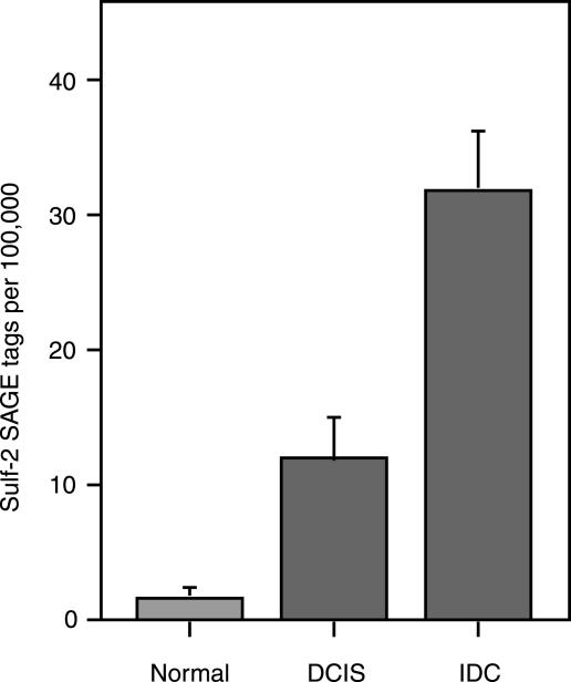
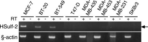
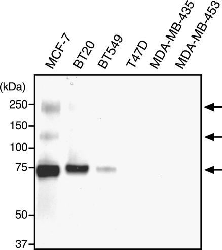
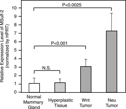
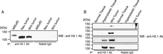
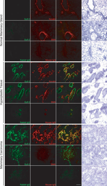
Sulf-2 is an endosulfatase with activity against glucosamine-6-sulfate modifications within subregions of intact heparin. The enzyme has the potential to modify the sulfation status of extracellular heparan sulfate proteoglycan (HSPG) glycosaminoglycan chains and thereby to regulate interactions with HSPG-binding proteins. In the present investigation, data mining from published studies was employed to establish Sulf-2 mRNA upregulation in human breast cancer. We further found that cultured breast carcinoma cells expressed Sulf-2 mRNA and released enzymatically active proteins into conditioned medium. In two mouse models of mammary carcinoma, Sulf-2 mRNA was upregulated in comparison to its expression in normal mammary gland. Although mRNA was present in normal tissues, Sulf-2 protein was undetectable; it was, however, detected in some premalignant lesions and in tumors. The protein was localized to the epithelial cells of the tumors. In support of the possible mechanistic relevance of Sulf-2 upregulation in tumors, purified recombinant Sulf-2 promoted angiogenesis in the chick chorioallantoic membrane assay.


Figures

Figure 1
Figure 1
SAGE analysis of Sulf-2 expression in human breast cancer. Data pertinent to Sulf-2 were mined from a published SAGE analysis of human breasts [23]. Sulf-2 tags were enumerated from 4 normal breast tissue libraries, 7 DCIS libraries, and 12 IDC libraries. The total number of SAGE tags in the analysis was 1.75 million. The incidences of Sulf-2 tags in the different tissue libraries were 0.0018% (normal), 0.012% (DCIS), and 0.032% (IDC). The graph shows the mean number of Sulf-2 tags per 100,000 total tags from the libraries for each class of tissue. The error bars indicate SEM. The probabilities provided in the figure and text are based on a t-test comparison in the source study [23].
Figure 2
Figure 2
Sulf-2 mRNA in human breast carcinoma cell lines. RT-PCR was performed on cDNA that were prepared from eight human breast carcinoma cell lines with primer pairs for Sulf-2 and β-actin primer. A 314-bp HSulf-2 cDNA product (indicated by an arrow) was amplified from MCF-7, BT20, and BT549 cells.
Figure 3
Figure 3
Sulf-2 in the CM of human breast carcinoma cell lines. The CM of the indicated breast carcinoma cell lines was collected, and Western blot analysis with a Sulf-2 antibody (H2.3) was performed. The apparent molecular mass of the major Sulf-2 species was 72 kDa. Minor species were evident at 135 and 240 kDa.
Figure 4
Figure 4
Arylsulfatase activity of Sulf-2 in MCF-7 CM. Different volumes of CM obtained from MCF-7 cells or HSulf-2–transfected CHO cells were incubated with Sulf-2 antibody (H2.3) or rabbit IgG coupled to protein A beads. Bead-bound material was tested for arylsulfatase activity against a 10-mM 4-MUS substrate. The same results were obtained from three different experiments. (▪) MCF-7 CM + Sulf-2 antibody (H2.3) beads; (□) HSulf-2 CHO CM + Sulf-2 antibody beads; (▴) MCF-7 CM + rabbit IgG beads; (△) HSulf-2 CHO CM + rabbit IgG beads.
Figure 5
Figure 5
Sulf-2 mRNA in MMTV-Wnt1 and MMTV-Neu transgenic mouse mammary carcinoma tissues. Real-time PCR was performed on cDNA prepared from normal mouse mammary glands (10 independent samples), hyperplastic tissues (9), and tumors from MMTV-Wnt1 (12) and MMTV-Neu (6) transgenic mice. Sulf-2 mRNA expression was normalized relative to HPRT expression. Error bars indicate SEM. Statistical significance was computer with Student's t test.
Figure 6
Figure 6
Sulf-2 expression in normal mouse tissues and mammary gland tumors. Detergent lysates of the indicated tissues were subjected to immunoprecipitation and Western blot analysis with a Sulf-2 (H2.1) antibody or normal rabbit IgG, as described in Materials and Methods section. (A) Analysis of expression in MMTV-Neu tumor, MMTV-Wnt1 tumor, and cell extracts of COS-7 cells transfected with MSulf-2. (B) Four sets of normal mammary gland from FVB/N mouse, and hyperplastic and tumor tissues from a littermate MMTV-Wnt1 transgenic mouse were subjected to immunoprecipitation/Western blot analysis, as described above. Sulf-2 was detected in all of the tumors, in two of four of the hyperplastic tissues, and in none of the normal tissue extracts.
Figure 7
Figure 7
Sulf-2 in the epithelia of hyperplastic mammary tissues and mammary tumors. Sections of mammary tumors and hyperplastic tissues from MMTV-Wnt1 mice and normal mammary glands from FVB/N mice were processed for dual-color immunocytochemistry and bright field histology. The left column shows staining with a Sulf-2 antibody (H2.1; green signal). The second column shows staining with anti–pan cytokeratin (keratin; epithelial cell marker) or anti–SMA (myoepithelial cell marker) antibodies (red signal). The third column shows the overlay of the two fluorescent images. The fourth column shows a bright field image with hematoxylin staining. Scale bar = 100 μm.
Figure 8
Figure 8
Angiogenic activity of Sulf-2 in the CAM assay. Ten-day-old chicken embryo CAMs were treated with purified Sulf-2, Sulf-2ΔCC, or VEGF in 50 mM HEPES (pH 8.0). Three days after the treatment, the number of vessel branch points was counted. Data were pooled from three independent experiments. Means and SEM are shown. Statistical comparisons were performed with Student's t test.

References

    1. Esko JD, Lindahl U. Molecular diversity of heparan sulfate. J Clin Invest. 2001;108:169–173. - PMC - PubMed
    1. Sasisekharan R, Shriver Z, Venkataraman G, Narayanasami U. Roles of heparan-sulphate glycosaminoglycans in cancer. Nat Rev Cancer. 2002;2:521–528. - PubMed
    1. Gallagher JT. Heparan sulfate: growth control with a restricted sequence menu. J Clin Invest. 2001;108:357–361. - PMC - PubMed
    1. Esko JD, Selleck SB. ORDER OUT OF CHAOS: assembly of ligand binding sites in heparan sulfate. Annu Rev Biochem. 2002;71:435–471. - PubMed
    1. Schlessinger J, Plotnikov AN, Ibrahimi OA, Eliseenkova AV, Yeh BK, Yayon A, Linhardt RJ, Mohammadi M. Crystal structure of a ternary FGF–FGFR–heparin complex reveals a dual role for heparin in FGFR binding and dimerization. Mol Cell. 2000;6:743–750. - PubMed

Publication types