Characterization of functional heme domains from soluble guanylate cyclase
- PMID: 16331987
- PMCID: PMC2572136
- DOI: 10.1021/bi051601b
Characterization of functional heme domains from soluble guanylate cyclase
Abstract
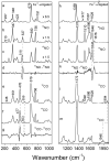
Soluble guanylate cyclase (sGC) is a heterodimeric, nitric oxide (NO)-sensing hemoprotein composed of two subunits, alpha1 and beta1. NO binds to the heme cofactor in the beta1 subunit, forming a five-coordinate NO complex that activates the enzyme several hundred-fold. In this paper, the heme domain has been localized to the N-terminal 194 residues of the beta1 subunit. This fragment represents the smallest construct of the beta1 subunit that retains the ligand-binding characteristics of the native enzyme, namely, tight affinity for NO and no observable binding of O(2). A functional heme domain from the rat beta2 subunit has been localized to the first 217 amino acids beta2(1-217). These proteins are approximately 40% identical to the rat beta1 heme domain and form five-coordinate, low-spin NO complexes and six-coordinate, low-spin CO complexes. Similar to sGC, these constructs have a weak Fe-His stretch [208 and 207 cm(-)(1) for beta1(1-194) and beta2(1-217), respectively]. beta2(1-217) forms a CO complex that is very similar to sGC and has a high nu(CO) stretching frequency at 1994 cm(-)(1). The autoxidation rate of beta1(1-194) was 0.073/min, while the beta2(1-217) was substantially more stable in the ferrous form with an autoxidation rate of 0.003/min at 37 degrees C. This paper has identified and characterized the minimum functional ligand-binding heme domain derived from sGC, providing key details toward a comprehensive characterization.
Figures


Similar articles
-
Probing domain interactions in soluble guanylate cyclase.Biochemistry. 2011 May 24;50(20):4281-90. doi: 10.1021/bi200341b. Epub 2011 May 3. Biochemistry. 2011. PMID: 21491957 Free PMC article.
-
Resonance raman characterization of the heme domain of soluble guanylate cyclase.Biochemistry. 1998 Nov 17;37(46):16289-97. doi: 10.1021/bi981547h. Biochemistry. 1998. PMID: 9819221
-
Identification of histidine 105 in the beta1 subunit of soluble guanylate cyclase as the heme proximal ligand.Biochemistry. 1998 Mar 31;37(13):4502-9. doi: 10.1021/bi972686m. Biochemistry. 1998. PMID: 9521770
-
Ligand specificity of H-NOX domains: from sGC to bacterial NO sensors.J Inorg Biochem. 2005 Apr;99(4):892-902. doi: 10.1016/j.jinorgbio.2004.12.016. J Inorg Biochem. 2005. PMID: 15811506 Review.
-
Structure and Activation of Soluble Guanylyl Cyclase, the Nitric Oxide Sensor.Antioxid Redox Signal. 2017 Jan 20;26(3):107-121. doi: 10.1089/ars.2016.6693. Epub 2016 Apr 26. Antioxid Redox Signal. 2017. PMID: 26979942 Free PMC article. Review.
Cited by
-
Smooth muscle cell CYB5R3 preserves cardiac and vascular function under chronic hypoxic stress.J Mol Cell Cardiol. 2022 Jan;162:72-80. doi: 10.1016/j.yjmcc.2021.09.005. Epub 2021 Sep 15. J Mol Cell Cardiol. 2022. PMID: 34536439 Free PMC article.
-
Insight into the rescue of oxidized soluble guanylate cyclase by the activator cinaciguat.Chembiochem. 2012 May 7;13(7):977-81. doi: 10.1002/cbic.201100809. Epub 2012 Mar 30. Chembiochem. 2012. PMID: 22474005 Free PMC article.
-
The Influence of Nitric Oxide on Soluble Guanylate Cyclase Regulation by Nucleotides: ROLE OF THE PSEUDOSYMMETRIC SITE.J Biol Chem. 2015 Jun 19;290(25):15570-15580. doi: 10.1074/jbc.M115.641431. Epub 2015 Apr 23. J Biol Chem. 2015. PMID: 25907555 Free PMC article.
-
Probing domain interactions in soluble guanylate cyclase.Biochemistry. 2011 May 24;50(20):4281-90. doi: 10.1021/bi200341b. Epub 2011 May 3. Biochemistry. 2011. PMID: 21491957 Free PMC article.
-
Regulation of sGC via hsp90, Cellular Heme, sGC Agonists, and NO: New Pathways and Clinical Perspectives.Antioxid Redox Signal. 2017 Feb 1;26(4):182-190. doi: 10.1089/ars.2016.6690. Epub 2016 May 2. Antioxid Redox Signal. 2017. PMID: 26983679 Free PMC article.
References
-
- Zhao Y, Marletta MA. Localization of the heme binding region of soluble guanylate cyclase. Biochemistry. 1997;36:15959–15964. - PubMed
-
- Stone JR, Marletta MA. Soluble Guanylate Cyclase from Bovine Lung: Activation with Nitric Oxide and Carbon Monoxide and Spectral Characterization of the Ferrous and Ferric States. Biochemistry. 1994;33:5636–5640. - PubMed
-
- Watmough NJ, Butland G, Cheesman MR, Moir JW, Richardson DJ, Spiro S. Nitric oxide in bacteria: synthesis and consumption. Biochim. Biophys. Acta. 1999;1411:456–474. - PubMed
Publication types
MeSH terms
Substances
Grants and funding
LinkOut - more resources
Full Text Sources
Other Literature Sources
Molecular Biology Databases
Research Materials

