. 2005 Dec 14;127(49):17412-20.
doi: 10.1021/ja055846n.

Uracil-directed ligand tethering: an efficient strategy for uracil DNA glycosylase (UNG) inhibitor development

Affiliations

Uracil-directed ligand tethering: an efficient strategy for uracil DNA glycosylase (UNG) inhibitor development

Yu Lin Jiang et al. J Am Chem Soc. .

Abstract

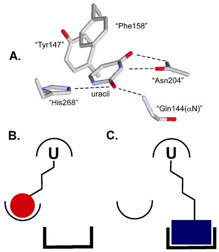
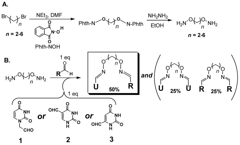
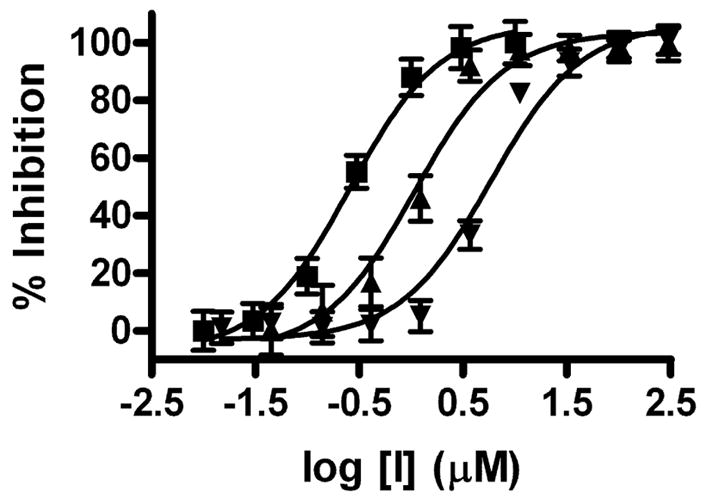
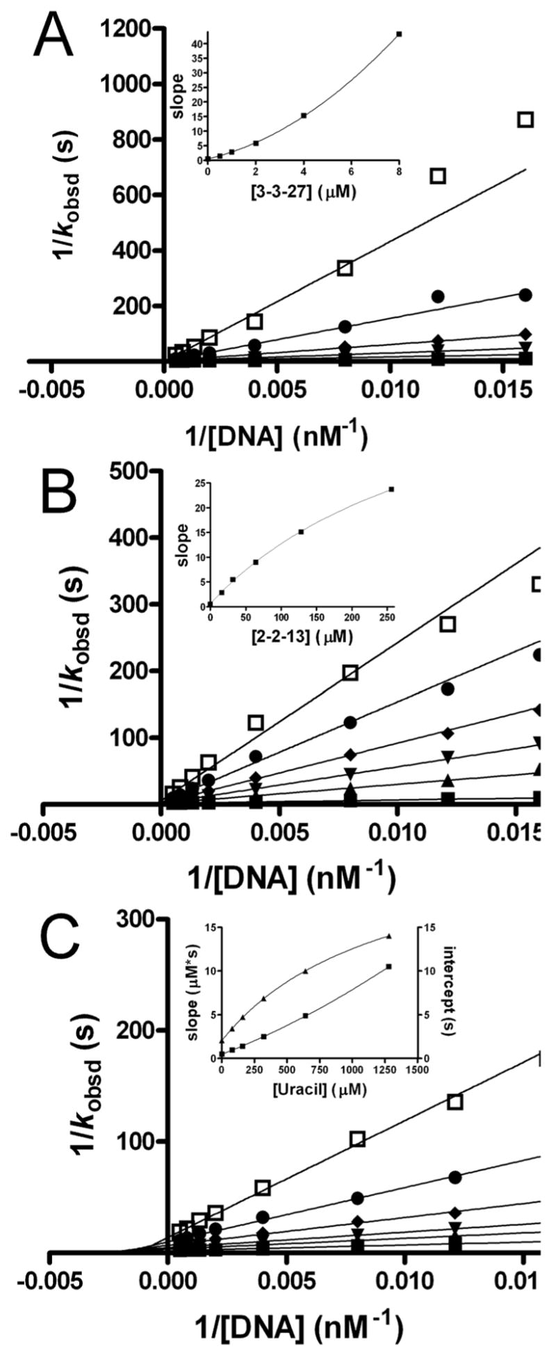
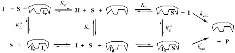
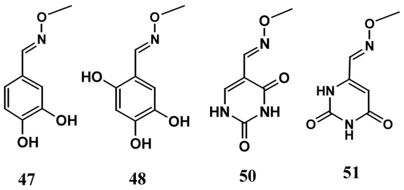
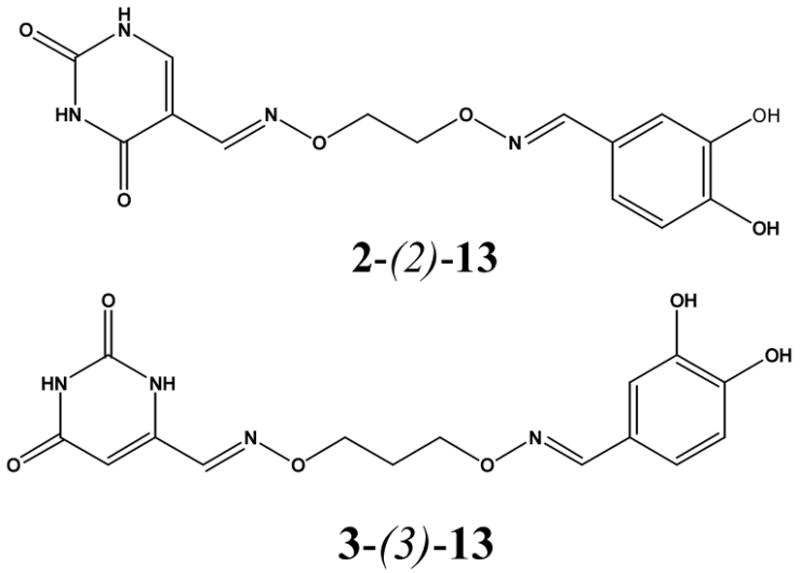
Uracil DNA glycosylase (UNG) is an important DNA repair enzyme that recognizes and excises uracil bases in DNA using an extrahelical recognition mechanism. It is emerging as a desirable target for small-molecule inhibitors given its key role in a wide range of biological processes including the generation of antibody diversity, DNA replication in a number of viruses, and the formation of DNA strand breaks during anticancer drug therapy. To accelerate the discovery of inhibitors of UNG we have developed a uracil-directed ligand tethering strategy. In this efficient approach, a uracil aldehyde ligand is tethered via alkyloxyamine linker chemistry to a diverse array of aldehyde binding elements. Thus, the mechanism of extrahelical recognition of the uracil ligand is exploited to target the UNG active site, and alkyloxyamine linker tethering is used to randomly explore peripheral binding pockets. Since no compound purification is required, this approach rapidly identified the first small-molecule inhibitors of human UNG with micromolar to submicromolar binding affinities. In a surprising result, these uracil-based ligands are found not only to bind to the active site but also to bind to a second uncompetitive site. The weaker uncompetitive site suggests the existence of a transient binding site for uracil during the multistep extrahelical recognition mechanism. This very general inhibitor design strategy can be easily adapted to target other enzymes that recognize nucleobases, including other DNA repair enzymes that recognize other types of extrahelical DNA bases.


Figures

Figure 1
Figure 1
Extrahelical binding of uracil to the UNG active site and the general strategy for uracil-directed ligand tethering. (A) Structure of UNG bound to uracil (pdb code 2eug). The residue numbering is for the human enzyme. (B) and (C) The uracil ligand (U) that targets the UDG active site is covalently tethered to two different ligands that can interact with distinct binding surfaces near the active site.
Figure 2
Figure 2
Synthesis of oxime libraries based on uracil and RCHO. (A) Synthesis of diaminoalkanediol tethers of variable length. (B) Construction of the uracil-oxime library based on the uracil aldehydes (13) and a series of aldehyde compounds (RCHO). The products consist of a 1:2:1 mixture of the heterodimer (U^R), and the two homodimers (U^U and R^R) connected via alkane linkers of lengths 2–6. One equivalent of total diaminoalkanediol is added to each reaction. Each linker length is present at one-fifth of the total concentration.
Figure 3
Figure 3
High-throughput (HTP) UDG kinetic assay. (A) The HTP assay relies on molecular beacon technology. Excision of multiple uracil bases by the enzyme destabilizes the hairpin structure thereby releasing the 5′ FAM fluorophore from the quenching effects of the 3′ dabsyl group. (B) Steady-state kinetic analysis of the hUDG reaction using the molecular beacon hairpin substrate.
Figure 4
Figure 4
Representative HTP screening results using the molecular beacon substrate. (A) Screen of oxime dimer mixtures derived from uracil aldehyde 1 and aryl aldehydes 1317. No inhibition was observed for any oxime derived from 1 regardless of linker length (n). (B) Screen of oxime dimer mixtures derived from uracil aldehyde 3 and aryl aldehydes 1317. The mixed oxime derived from 3 and 13 shows significant inhibition and this derivative was further optimized. For 1417, the observed inhibition represents that from the 3-3 homodimers that are present in the mixtures.
Figure 5
Figure 5
IC50 analysis of for 2-(2)-13 (▼), 3-(3)-13 (▲) and 3-(3)-27 (■).
Figure 6
Figure 6
Mode of inhibition analysis. Double reciprocal plots and secondary slope and intercept replots for inhibition by increasing concentrations of (A) 3-(3)-27, (B) 2-(2)-13, and (C) uracil. Slope and intercept effects in the inset to (C) are shown as squares and triangles, respectively.
Scheme 1
Scheme 1
Inhibition Mechanisms for 3-(3)-27 and 2-(2)-13 and Uracila aOnly 3-(3)-27, 2-(2)-13 and uracil have mechanisms that include the kcat′ step. The mechanism for 3-(3)-and uracil do not include the equilibrium constant Kn, and the mechanism for 2-(2)-13 does not include the equilibria Kc or Knc.
Scheme 2
Scheme 2
O-methyl Oxime Derivatives of the Aldehyde Binding Elements of 2-(2)-13 and 3-(3)-27
Chart 1
Chart 1
Heterodimer Oximes Identified from Deconvolution of Active Mixtures

References

    1. Lindahl T, Wood RD. Science. 1999;286:1897–1905. - PubMed
    1. Di Noia J, Neuberger MS. Nature. 2002;419:43–48. - PubMed
    1. Imai K, Slupphaug G, Lee WI, Revy P, Nonoyama S, Catalan N, Yel L, Forveille M, Kavli B, Krokan HE, Ochs HD, Fischer A, Durandy A. Nature Immunol. 2003;4:1023–1028. - PubMed
    1. Storb U, Stavnezer J. Curr Biol. 2002;12:R725–727. - PubMed
    1. Chen R, Wang H, Mansky LM. J Gen Virol. 2002;83:2339–2345. - PubMed

Publication types