. 2005 Sep-Oct;22(5):685-91.
doi: 10.1017/S0952523805225178.

Optical imaging of the intrinsic signal as a measure of cortical plasticity in the mouse

Affiliations

Optical imaging of the intrinsic signal as a measure of cortical plasticity in the mouse

Jianhua Cang et al. Vis Neurosci. 2005 Sep-Oct.

Abstract

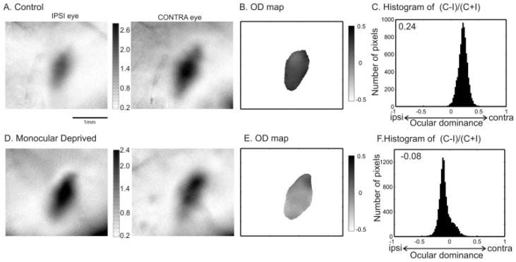
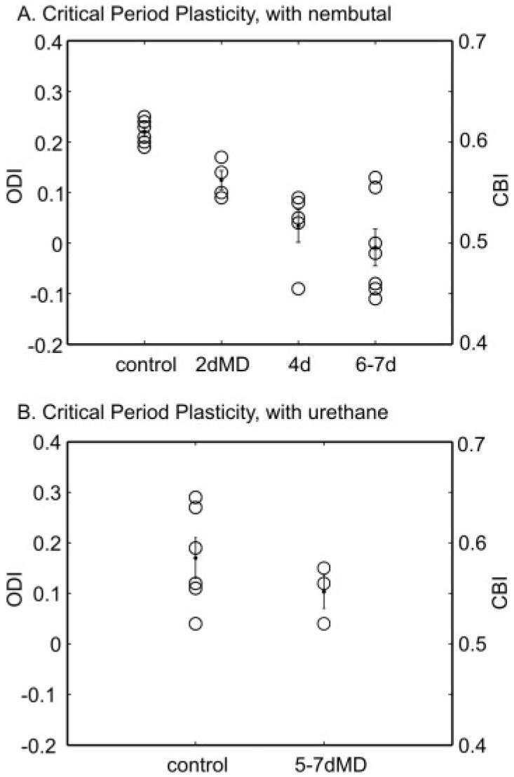
The responses of cells in the visual cortex to stimulation of the two eyes changes dramatically following a period of monocular visual deprivation (MD) during a critical period in early life. This phenomenon, referred to as ocular dominance (OD) plasticity, is a widespread model for understanding cortical plasticity. In this study, we designed stimulus patterns and quantification methods to analyze OD in the mouse visual cortex using optical imaging of intrinsic signals. Using periodically drifting bars restricted to the binocular portion of the visual field, we obtained cortical maps for both contralateral (C) and ipsilateral (I) eyes and computed OD maps as (C - I)/(C + I). We defined the OD index (ODI) for individual animals as the mean of the OD map. The ODI obtained from an imaging session of less than 30 min gives reliable measures of OD for both normal and monocularly deprived mice under Nembutal anesthesia. Surprisingly, urethane anesthesia, which yields excellent topographic maps, did not produce consistent OD findings. Normal Nembutal-anesthetized mice have positive ODI (0.22 +/- 0.01), confirming a contralateral bias in the binocular zone. For mice monocularly deprived during the critical period, the ODI of the cortex contralateral to the deprived eye shifted negatively towards the nondeprived, ipsilateral eye (ODI after 2-day MD: 0.12 +/- 0.02, 4-day: 0.03 +/- 0.03, and 6- to 7-day MD: -0.01 +/- 0.04). The ODI shift induced by 4-day MD appeared to be near maximal, consistent with previous findings using single-unit recordings. We have thus established optical imaging of intrinsic signals as a fast and reliable screening method to study OD plasticity in the mouse.


Figures

Fig. 1
Fig. 1
Visual stimulus paradigm for imaging ocular dominance of cortical responses. (A) Monitor placement and stimulus patterns used to stimulate both ipsilateral and contralateral eyes for imaging OD. The monitor (40 cm × 30 cm) was placed 25 cm in front of the mouse, with its midline (defined as 0 deg, marked by a cross) aligned to the midline of the animal. Drifting thin bars (2 deg wide, spatial frequency of 1 cycle/80 deg, and temporal frequency of 1 cycle/6-8 s) were displayed in the binocular visual field (marked by dotted lines, -5 deg to 15 deg on the monitor) to assess OD. (B) Iso-azimuth (Solid lines) and iso-elevation (dashed lines) obtained from full-screen stimulation through the contralateral eye. The spacing of the contour lines is 2.5 deg. The scale bar applies to C and D as well. (C) Cortical response magnitude to the restricted stimuli through the ipsilateral eye (left eye). (D) Response through the contralateral eye. The gray scale shown to the right is used to visualize the response magnitude as fractional change in reflection ×104.
Fig. 2
Fig. 2
Quantification of cortical responses to determine OD. (A) Responsive area of the ipsilateral eye magnitude map shown in Fig. 1. The area was selected by smoothing the ipsilateral eye map with a low-pass filter and then thresholding it at 30% of peak response amplitude. The nonsmoothed magnitude of the pixels within the responsive area is shown (gray scale shown to the right). (B) Response magnitude map of the contralateral eye of the same responsive area as in A shown in the same gray scale. (C) OD map in the region of interest. The OD score was calculated as (C - I)/(C + I) for every responsive pixel and plotted in gray scale, with C and I representing the non-smoothed response magnitudes of each pixel from the contralateral and ipsilateral eye maps, respectively. (D) Histogram of the OD scores for all responsive pixels. The average of (C - I)/(C + I) for all pixels is 0.29, confirming a strong contralateral bias in the binocular zone of the visual cortex in this adult mouse. The scale bar applies to all the panels showing maps (A, B, & C).
Fig. 3
Fig. 3
Imaging OD plasticity during the critical period. The map of response magnitudes to the ipsilateral and contralateral eyes are shown for a control mouse (P30) in A and for a monocularly deprived mouse (MD from P28 to P35) in D. The 2-D OD maps of the two mice are computed and plotted in B and E, respectively. Figs. C and F show the histograms of the OD scores obtained from nonsmoothed maps. The ODI is 0.24 for the control mouse, indicating a strong contralateral bias; -0.08 for the MD mouse, showing a slight bias towards the nondeprived, ipsilateral eye. The scale bar applies to all the panels showing maps (A, B, D, & E).
Fig. 4
Fig. 4
Effect of MD duration on OD shift revealed by imaging. (A) The ODI of each animal (open circle) is plotted against the duration of MD which started between P26 and P28. The mean and SEM of each group are also shown (dots).The ODI is related to the CBI used for analyzing single unit data by CBI = (ODI + 1)/2, so the corresponding CBI is also shown as the right axis for this and the following figures. Four-day MD induced a significant shift in ODI towards to the nondeprived eye (Control: 0.22 ± 0.01 vs. 4-day: 0.03 ± 0.03, P value < 0.05). The shift caused by 4-day MD is not significantly different from that by deprivation longer than 6 days (-0.01 ± 0.04, P > 0.05). In addition, 2-day MD did not induce a significant shift of the ODI (2-day: 0.12 ± 0.02, P > 0.05). (B) Same plot for results obtained from experiments using urethane as anesthesia. The ODI calculated from the imaging data is not significantly different between the control (0.17 ± 0.04, n = 6) and MD group (0.10 ± 0.03, n = 3; P value = 0.32).

Similar articles

Cited by

References

    1. Cancedda L, Putignano E, Impey S, Maffei L, Ratto GM, Pizzorusso T. Patterned vision causes CRE-mediated gene expression in the visual cortex through PKA and ERK. Journal of Neuroscience. 2003;23:7012–7020. - PMC - PubMed
    1. Fagiolini M, Fritschy JM, Low K, Mohler H, Rudolph U, Hensch TK. Specific GABAA circuits for visual cortical plasticity. Science. 2004;303:1681–1683. - PubMed
    1. Fagiolini M, Hensch TK. Inhibitory threshold for critical period activation in primary visual cortex. Nature. 2000;404:183–186. - PubMed
    1. Gianfranceschi L, Siciliano R, Walls J, Morales B, Kirkwood A, Huang ZJ, Tonegawa S, Maffei L. Visual cortex is rescued from the effects of dark rearing by overexpression of BDNF. Proceedings of the National Academy of Sciences of the U.S.A. 2003;100:12486–12491. - PMC - PubMed
    1. Gordon JA, Stryker MP. Experience-dependent plasticity of binocular responses in the primary visual cortex of the mouse. Journal of Neuroscience. 1996;16:3274–3286. - PMC - PubMed

Publication types