

In vitro hypoxia-conditioned colon cancer cell lines derived from HCT116 and HT29 exhibit altered apoptosis susceptibility and a more angiogenic profile in vivo
- PMID: 16333244
- PMCID: PMC2361533
- DOI: 10.1038/sj.bjc.6602864
In vitro hypoxia-conditioned colon cancer cell lines derived from HCT116 and HT29 exhibit altered apoptosis susceptibility and a more angiogenic profile in vivo
Abstract
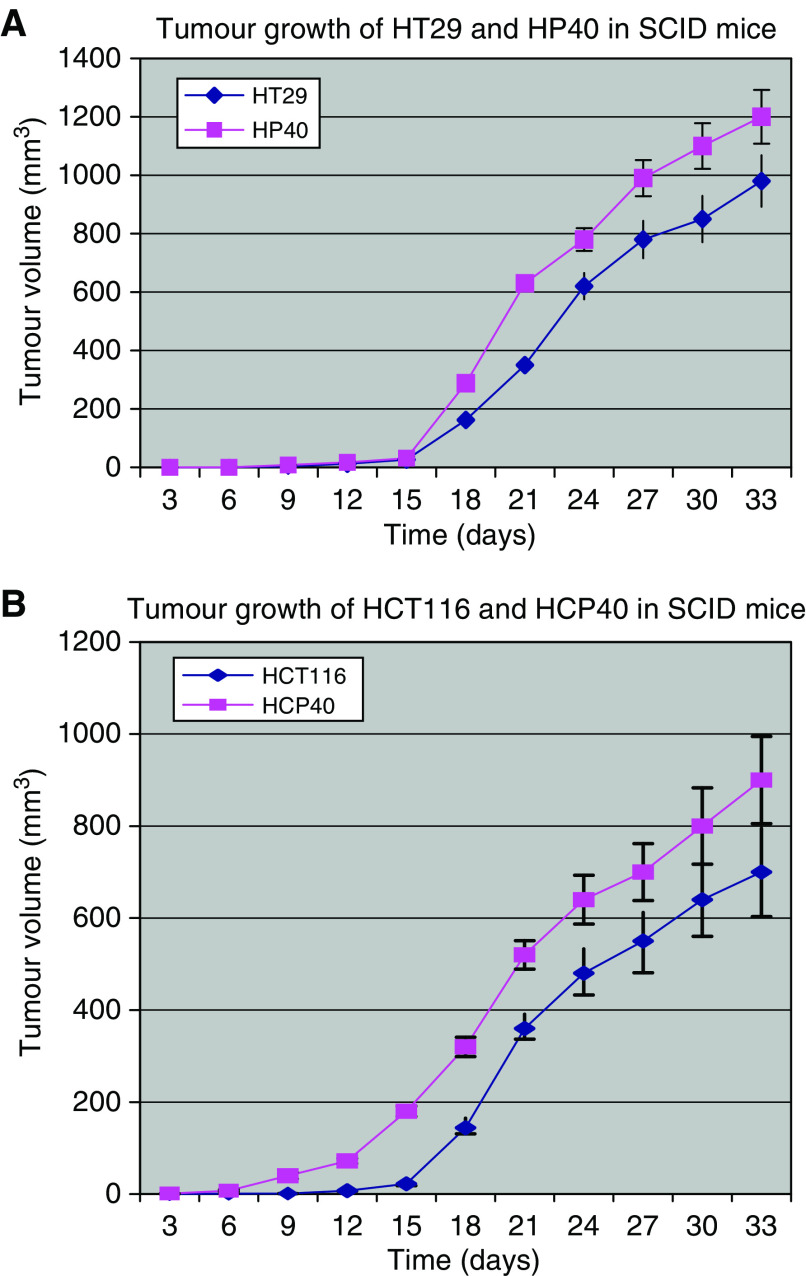
Hypoxia is an important selective force in the clonal evolution of tumours. Through HIF-1 and other transcription factors combined with tumour-specific genetic alterations, hypoxia is a dominant factor in the angiogenic phenotype. Cellular adaptation to hypoxia is an important requirement of tumour progression independent of angiogenesis. The adaptive changes, insofar as they alter hypoxia-induced apoptosis, are likely to determine responsiveness to antiangiogenic strategies. To investigate this adaptation of tumour cells to hypoxia, we recreated in vitro the in vivo situation of chronic intermittent exposure to low-oxygen levels. The colon carcinoma cell lines HT29 and HCT116 were subjected to 40 episodes of sublethal hypoxia (4 h) three times a week. The resulting two hypoxia-conditioned cell lines have been maintained in culture for more than 2 years. In both cell lines changes in doubling times occurred: in HT29 an increase, and in HCT116 a decrease. Cell survival in response to hypoxia and to DNA damage differed strikingly in the two cell lines. The HT29 hypoxia-conditioned cells were more resistant than the parental line to a 24 h hypoxic challenge, while those from HCT116 surprisingly were more sensitive. Sensitivity to cisplatin in vitro was also significantly different for the hypoxia-conditioned compared with the parental lines, suggesting a change in pathways leading to apoptosis following DNA damage signaling. The growth of both conditioned cell lines in vivo as xenografts in immunodeficient (SCID) mice was more rapid than their parental lines, and was accompanied in each by evidence of enhanced vascular proliferation as a consequence of the hypoxia-conditioning. Thus the changes in apoptotic susceptibility were independent of altered angiogenesis. The derivation of these lines provides a model for events within hypoxic regions of colon cancers, and for the acquisition of resistance and sensitivity characteristics that may have therapeutic implications for the use of antiangiogenesis drugs.
Figures


References
-
- Amellem O, Stokke T, Sandvik JA, Smedshammer L, Pettersen EO (1997) Hypoxia-induced apoptosis in human cells with normal p53 status and function, without any alteration in the nuclear protein level. Exp Cell Res 232: 361–370 - PubMed
-
- Brizel DM, Sibley GS, Prosnitz LR, Scher RL, Dewhirst MW (1997) Tumor hypoxia adversely effects the prognosis of carcinoma of the head and neck. Int J Radiat Oncol Biol Phys 38: 285–289 - PubMed
-
- Brown JM, Koong A (1991) Therapeutic advantage of hypoxic cells in tumors: a theoretical study. J Natl Cancer Inst 83: 178–185 - PubMed
-
- Brown JM (1999) The hypoxic cell: a target for selective cancer therapy – eighteenth Bruce F. Cain Memorial Award lecture. Cancer Res 59: 5863–5870 - PubMed
Publication types
MeSH terms
Grants and funding
LinkOut - more resources
Full Text Sources
