. 2005 Winter;10(4):340-8.
doi: 10.1379/csc-98r1.1.

Heat shock protein 70, heat shock protein 32, and vascular endothelial growth factor production and their effects on lipopolysaccharide-induced apoptosis in porcine aortic endothelial cells

Affiliations

Heat shock protein 70, heat shock protein 32, and vascular endothelial growth factor production and their effects on lipopolysaccharide-induced apoptosis in porcine aortic endothelial cells

Chiara Bernardini et al. Cell Stress Chaperones. 2005 Winter.

Abstract

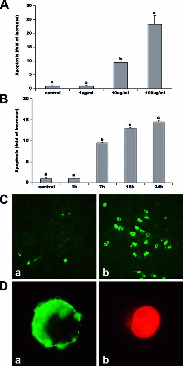
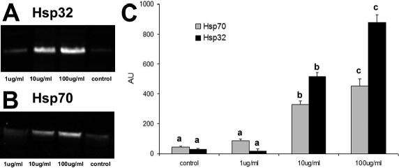
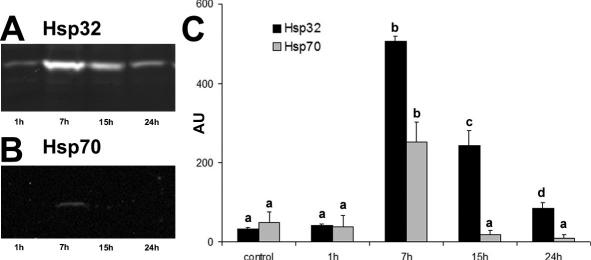
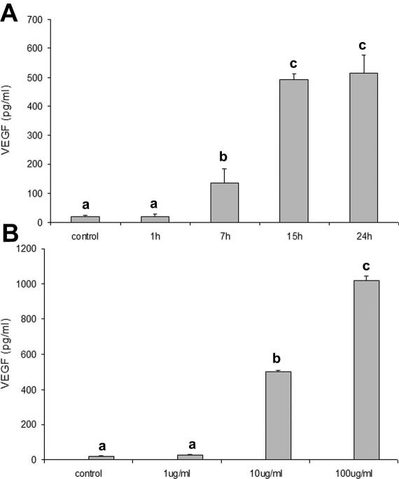
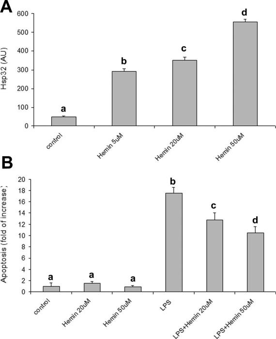
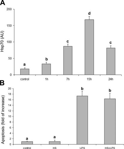
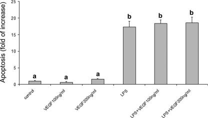
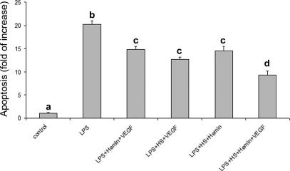
Lipopolysaccharide (LPS) is a highly proactive molecule that causes in vivo a systemic inflammatory response syndrome and activates in vitro the inflammatory pathway in different cellular types, including endothelial cells (EC). Because the proinflammatory status could lead to EC injury and apoptosis, the expression of proinflammatory genes must be finely regulated through the induction of protective genes. This study aimed at determining whether an LPS exposure is effective in inducing apoptosis in primary cultures of porcine aortic endothelial cells and in stimulating heat shock protein (Hsp)70 and Hsp32 production as well as vascular endothelial growth factor (VEGF) secretion. Cells between third and eighth passage were exposed to 10 microg/mL LPS for 1, 7, 15, and 24 hours (time-course experiments) or to 1, 10, and 100 microg/mL LPS for 7 and 15 hours (dose-response experiments). Apoptosis was not affected by 1 microg/mL LPS but significantly increased in a dose-dependent manner with the highest LPS doses. Furthermore, apoptosis rate increased only till 15 hours of LPS exposure. LPS stimulated VEGF secretion in a dose-dependent manner; its effect became significant after 7 hours and reached a plateau after 15 hours. Both Hsp70 and Hsp32 expressions were induced by LPS in a dose-dependent manner after 7 hours. Subsequent studies were addressed to evaluate the protective role of Hsp32, Hsp70, and VEGF. Hemin, an Hsp32 inducer (5, 20, 50 microM), and recombinant VEGF (100 and 200 ng/mL), were added to the culture 2 hours before LPS (10 microg/mL for 24 hours); to induce Hsp70 expression, cells were heat shocked (42 degrees C for 1 hour) 15 hours before LPS (10 microg/mL for 24 hours). Hemin exposure upregulated Hsp32 expression in a dose-dependent manner and protected cells against LPS-induced apoptosis. Heat shock (HS) stimulated Hsp70 expression but failed to reduce LPS-induced apoptosis; VEGF addition did not protect cells against LPS-induced apoptosis at any dose tested. Nevertheless, when treatments were associated, a reduction of LPS-induced apoptosis was always observed; the reduction was maximal when all the treatments (HS + Hemin + VEGF) were associated. In conclusion, this study demonstrates that LPS is effective in evoking "the heat shock response" with an increase of nonspecific protective molecules (namely Hsp70 and Hsp32) and of VEGF, a specific EC growth factor. The protective role of Hsp32 was also demonstrated. Further investigations are required to clarify the synergic effect of Hsp32, Hsp70, and VEGF, thus elucidating the possible interaction between these molecules.


Figures

Fig 1.
Fig 1.
LPS-induced apoptosis in PAEC. Apoptosis was assessed by 3 methods: the sandwich ELISA for histone-associated DNA fragments, the Vybrant Apoptosis Assay Kit, and the TUNEL method. Apoptosis is represented as fold of increase vs control on the basis of ELISA results. Apoptosis after exposure for 7 hours to different LPS doses (A) and after exposure to 10 μg/mL LPS for different periods (B). Data represent the mean ± SEM of 6 replicates. Different letters indicate significant differences (P < 0.001). (C) Representative TUNEL: cells with green fluorescence represent apoptotic cells. (a) control; (b) 24 hours of LPS. (D) Representative Annexin test: membranes of apoptotic cells showed a strong green labeling (a), whereas necrotic cells present both weak membrane Annexin V and strong nuclear propidium staining (b). ELISA, enzyme-linked immunosorbent assay; LPS, lipopolysaccharide; PAEC, porcine aortic endothelial cells
Fig 2.
Fig 2.
Hsp32 and Hsp70 expression in PAEC treated with increasing doses (1, 10, 100 μg/mL) of LPS for 7 hours. Representative Western blot of Hsp32 (A), Hsp70 (B), and relative Hsp70 and Hsp32 content (AU, Arbitrary Units) (C). Data represent the mean ± SEM of 3 replicates. Different letters indicate significant differences (P < 0.05). Hsp, heat shock protein; LPS, lipopolysaccharide; PAEC, porcine aortic endothelial cells
Fig 3.
Fig 3.
Hsp32 and Hsp70 expression in PAEC exposed to 10 μg/ mL LPS for different periods (1, 7, 15, and 24 hours). Representative Western blot of Hsp32 (A), Hsp70 (B), and relative Hsp70 and Hsp32 content (AU, Arbitrary Units) (C). Data represent the mean ± SEM of 3 replicates. Different letters indicate significant differences (P < 0.05). Hsp, heat shock protein; LPS, lipopolysaccharide; PAEC, porcine aortic endothelial cells
Fig 4.
Fig 4.
Effects of LPS on VEGF secretion by PAEC. (A) Effects of different periods of exposure to 10 μg/mL LPS. (B) Effects of 7 hours exposure to different LPS doses. Data represent the mean ± SEM of 3 replicates. Different letters indicate significant differences (P < 0.05). LPS, lipopolysaccharide; PAEC, porcine aortic endothelial cells; VEGF, vascular endothelial growth factor
Fig 5.
Fig 5.
Effects of hemin on Hsp32 expression and on LPS-induced apoptosis. (A) Hemin (5, 20, or 50 μM) upregulates Hsp32 expression after 15 hours exposure. Data represent the mean ± SEM of 3 replicates. Different letters indicate significant differences (P < 0.05). (B) Hemin reduces LPS-induced apoptosis. Apoptosis is represented as fold of increase vs control on the basis of the sandwich ELISA for histone-associated DNA fragments. Hemin (20 or 50 μM) was added to the culture medium 2 hours before LPS exposure (10 μg/mL for 24 hours). Data represent the mean ± SEM of 6 replicates. Different letters indicate significant differences (P < 0.001). ELISA, enzyme-linked immunosorbent assay; Hsp, heat shock protein; LPS, lipopolysaccharide
Fig 6.
Fig 6.
Effect of (HS) treatment on Hsp70 expression and LPS-induced apoptosis. (A) Heat shock stimulates Hsp70 expression. The cells were heat shocked (HS = 42°C for 1 hour) and recovered at different times (1, 7, 15, 24 hours). Data represent the mean ± SEM of 3 replicates. Different letters indicate significant differences (P < 0.05). (B) Hsp70 stimulation fails to reduce LPS-induced apoptosis. Apoptosis is represented as fold of increase vs control on the basis of the sandwich ELISA for histone-associated DNA fragments. The cells were heat shocked (HS = 42°C for 1 hour) and then cultured in standard conditions for 15 hours before LPS exposure (10 μg/mL for 24 hours). Data represent the mean ± SEM of 6 replicates. Different letters indicate significant differences (P < 0.001). ELISA, enzyme-linked immunosorbent assay; HS, heat shock; Hsp, heat shock protein; LPS, lipopolysaccharide
Fig 7.
Fig 7.
Effect of recombinant VEGF on LPS-induced apoptosis. VEGF fails to reduce LPS-induced apoptosis. Apoptosis is represented as fold of increase vs control on the basis of the sandwich ELISA for histone-associated DNA fragments. Recombinant VEGF (100 or 200 ng/mL) was added to the culture media 2 hours before LPS exposure (10 μg/mL for 24 hours). Data represent the mean ± SEM of 6 replicates. Different letters indicate significant differences (P < 0.001). ELISA, enzyme-linked immunosorbent assay; LPS, lipopolysaccharide; VEGF, vascular endothelial growth factor
Fig 8.
Fig 8.
Effect of treatment association on LPS-induced apoptosis. The maximum protective effect was obtained by the association of the 3 treatments (VEGF, hemin, heat shock). Apoptosis is represented as fold of increase vs control on the basis of the sandwich ELISA for histone-associated DNA fragments. Data represent the mean ± SEM of 6 replicates. Different letters indicate significant differences (P < 0.001). ELISA, enzyme-linked immunosorbent assay; LPS, lipopolysaccharide; VEGF, vascular endothelial growth factor

References

    1. Alon T, Hemo I, Itin A, Pe'er J, Stone J, Keshet E. Vascular endothelial growth factor acts as a survival factor for newly formed retinal vessels and has implications for retinopathy of prematurity. Nat Med. 1995;1:1024–1028.1078-8956(1995)001[1024:VEGFAA]2.0.CO;2 - PubMed
    1. Bach FH, Hancock WW, Ferran C. Protective genes expressed in endothelial cells: a regulatory response to injury. Immunol Today. 1997;18:483–486.0167-5699(1997)018[0483:PGEIEC]2.0.CO;2 - PubMed
    1. Bannerman DD, Goldblum SE. Mechanisms of bacterial lipopolysaccharide-induced endothelial apoptosis. Am J Physiol Lung Cell Mol Physiol. 2003;284:L899–L914.1040-0605(2003)284[L899:MOBLEA]2.0.CO;2 - PubMed
    1. Blann AD. Endothelial cell activation, injury, damage and dysfuction: separate entities or mutual terms? Blood Coag Fibrynolysis. 2000;11:623–630.0957-5235(2000)011[0623:ECAIDA]2.0.CO;2 - PubMed
    1. Bulger EM, Garcia I, Maier RV. Induction of heme-oxygenase 1 inhibits endothelial cell activation by endotoxin and oxidant stress. Surgery. 2003;134:146–152.0039-6060(2003)134[0146:IOHIEC]2.0.CO;2 - PubMed

Publication types

MeSH terms

Substances

LinkOut - more resources