. 1992;2(2):111-26.

The human skeletal alpha-actin gene is regulated by a muscle-specific enhancer that binds three nuclear factors

Affiliations

The human skeletal alpha-actin gene is regulated by a muscle-specific enhancer that binds three nuclear factors

G E Muscat et al. Gene Expr. 1992.

Abstract

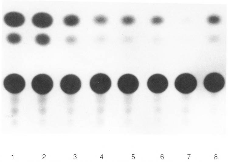
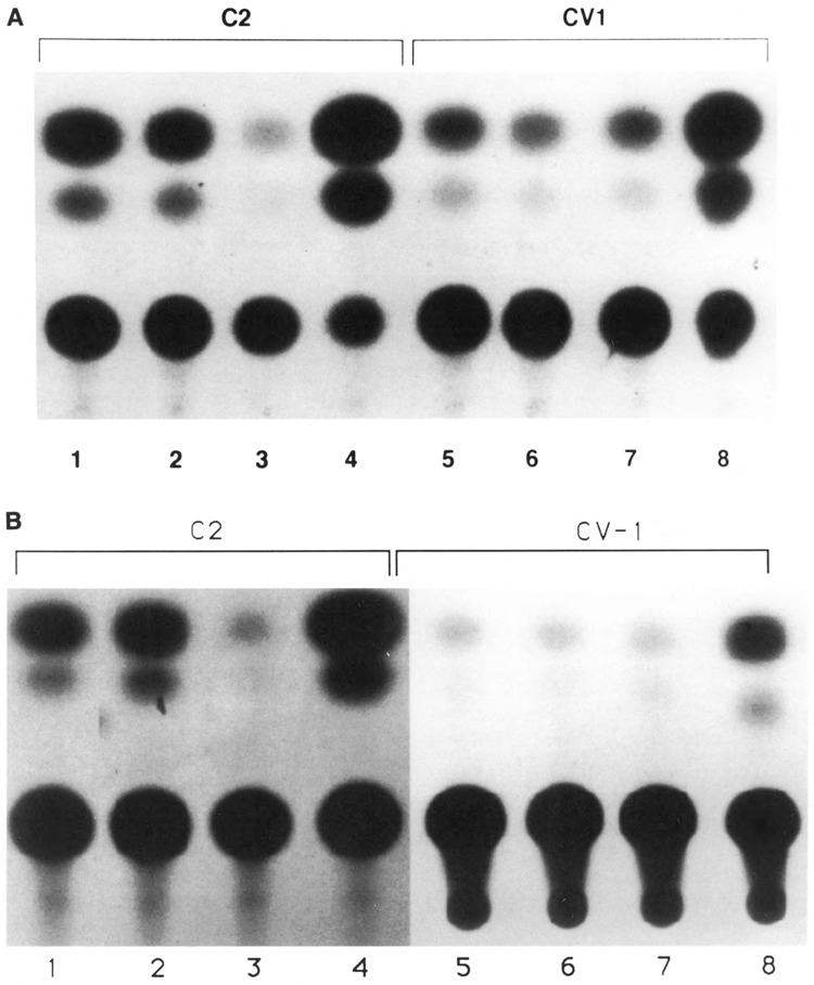
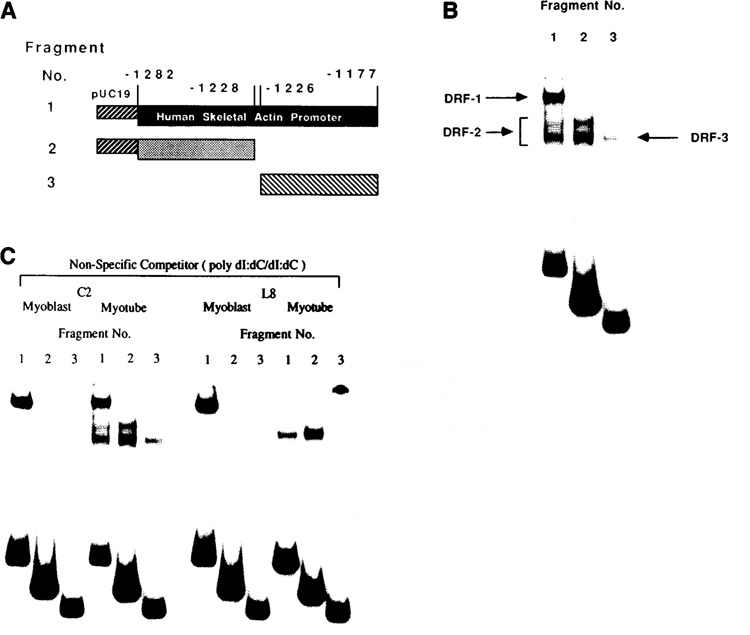
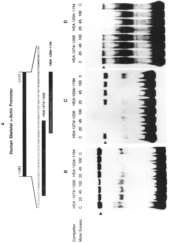
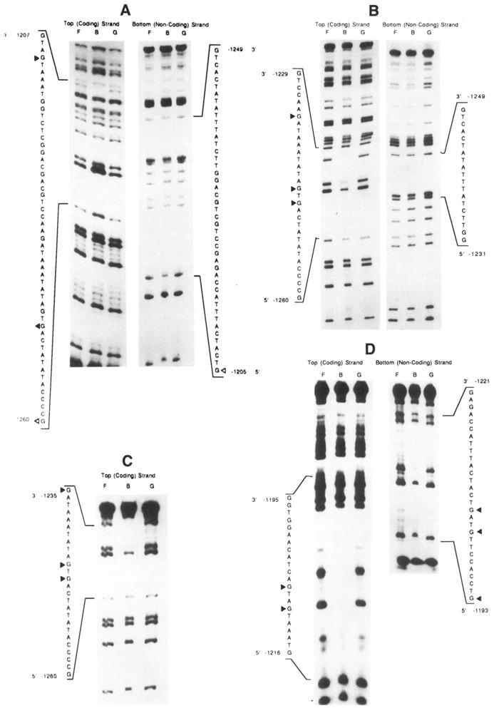
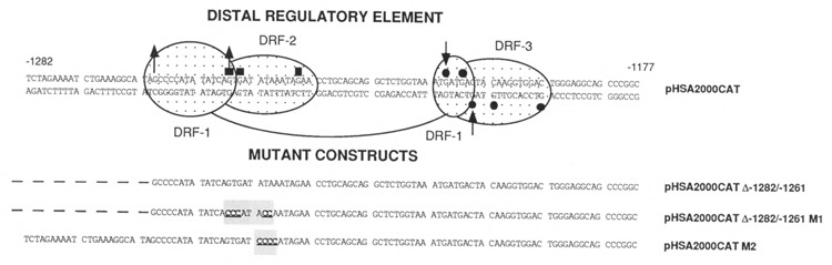
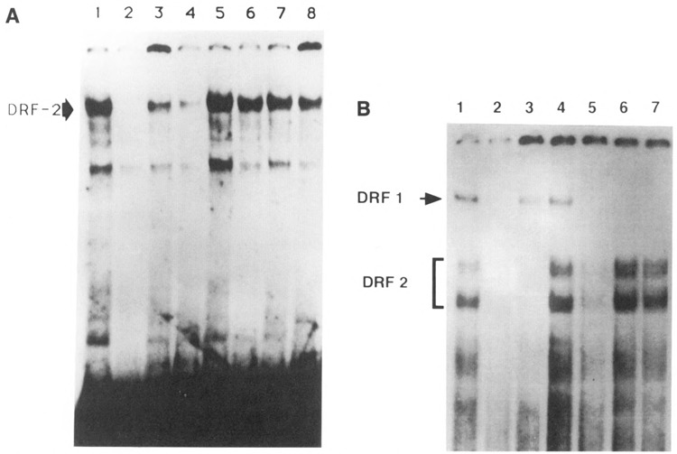
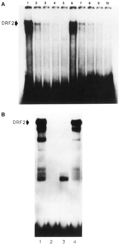
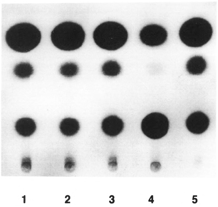
The tissue-specific distal promoter of the human skeletal alpha-actin gene (-1282 to -708) induces transcription in myogenic cells approximately 10-fold and, with the most proximal promoter domain (-153 to -87), it synergistically increases transcription 100-fold (Muscat and Kedes 1987). We report here that it is a short fragment of the distal promoter, the distal regulatory element (DRE) from -1282 to -1177 that functions as a muscle-specific, composite enhancer. An internal deletion in the DRE (delta -1282/-1151) in the context of the full-length 2000 bp promoter, resulted in a 10-fold reduction in transcription. Three distinct nuclear proteins, DRF-1, DRF-2, and DRF-3, interact specifically with the DRE between positions -1260 and -1193. A site specific mutation that abolishes DRF-2 binding also results in a 10-fold reduction in transcriptional activity. The DRF-2 nuclear protein has characteristics similar to those of the muscle-specific regulatory factor, MEF-2 (Buskin and Hauschka 1989; Gossett et al., 1989). Like the MEF-2 binding site in the muscle creatine kinase enhancer, the critical DRF-2 binding site is also an A/T-rich sequence element. The DRF-2 nuclear protein binds equally well to the MCK MEF-2 binding site and to the A/T-rich regulatory element of the skeletal muscle fast-twitch troponin C gene (Gahlmann and Kedes 1990). Furthermore, this troponin C site competes in vivo for DRF-2 driven expression of the skeletal alpha-actin gene in C2 cells. The DRF-2 site alone, however, does not activate transcription in muscle cells when linked to the SV40 promoter. We conclude that the DRF-2 binding element is a MEF-2 binding site that is required but insufficient for regulation of muscle-specific skeletal alpha-actin gene expression by the DRE. Thus, muscle-specific regulation of the human skeletal alpha-actin gene appears to require interactions between the other elements of the composite DRE enhancer with the protein:DNA complex formed by DRF-2.


Figures

Figure 1
Figure 1
CAT assays demonstrating the effect of internal deletions within the wild-type skeletal α-actin promoter on expression in C2 myotubes. Cells were transfected and harvested for CAT assays as described in Materials and Methods. Percentage of acetylation is shown in parenthesis. Lane 1: pHSA2000CAT (12.8%); lane 2: pHSA2000CAT LC (18.3%); lanes 3 (6.4%), 4 (2.2%), and 5 (2.6%): pHSA2000CAT Δ−1282/−1151; lanes 6 (2.8%), 7 (0.9%), and 8 (3.2%): pHSA2000CAT Δ−1282/−1089.
Figure 2
Figure 2
The distal regulatory region (DRE) is a muscle-specific enhancer. The element −1282/−1177 was cloned into the pCAT-promoter (Promega) upstream (Bgl II site: A, lanes 1, 2, 5, and 6) and downstream (Sph I site: B, lanes 1, 2, 5, and 6) of the CAT gene in sense (lanes 1 and 5) and antisense (lanes 2 and 6) orientation. CV-1 cells were transfected at 60% confluency, and expression was tested 48 hours later. In C2 myotubes the enhancer gave significant levels of activity above the pCAT plasmid (lanes 3 and 7) independent of location or orientation, but in non-muscle cells (CV-1 cells) no enhancer-like activity was detected. (β-actin served as a positive control (lanes 4 and 8). Reproductions of the original autoradiograms were obtained by digital scanning and photographed with a Presentation Technologies Montage system on a Macintosh Ilex computer with 8 megabytes of RAM.
Figure 3
Figure 3
A. Schematic representation of the three DNA fragments used in the electrophoretic mobility shift analysis of the distal regulatory region. The DNA fragments 1,2, and 3 contain skeletal α-actin sequences shown, plus pUC19 sequences including the Hind III/ Xba I region from the polylinker. B. Gel electrophoresis mobility shift analysis of the distal regulatory region. The DNA probes 1,2, and 3 were incubated with 5–10 μg of C2 myotube nuclear extract and assayed as described in Materials and Methods. Distal regulatory factors (DRF)-l, -2, and -3 refer to the three different factor complexes that interact with these DNA segments. C. The binding activities of DRF-1, -2, and -3 are differentially regulated in C2 and L8 cells during myogenesis. Each probe denoted by fragments 1,2, and 3 was incubated with nuclear extracts from C2 or L8 myoblasts and myotubes in the presence of poly(dl-dC)-poly(dl-dC) as the non-specific competitor. Fragment 1 interacts with DRF-1, DRF-2, and DRF-3. Fragment 2 interacts only with DRF-2, whereas Fragment 3 interacts only with DRF-3. C2 myotube lanes are the same as shown in Figure 3B to facilitate comparisons. The slow mobility band in lane 3, L8 myotubes, is radiolabeled material at the top of the gel well.
Figure 4
Figure 4
Gel electrophoresis mobility shift analysis of DRF-1, -2, and -3. A. The sequence of HSA−1282/−1177 is displayed, and the oligonucleotides used in competition (HSA−1274/−1226 and HSA−1234/−1194) are represented by the cross-hatched rectangular boxes. B. The effect of competition by the two oligonucleotides on the complex DRF-1 formed with the DRE probe in C2 myoblast nuclear extracts. Poly(dI-dC)poly(dI-dC) was used as a non-specific competitor. C. The effect of competition by the two oligonucleotides on the complex DRF-2, formed with the DRE (HSA−1282/−1177) in C2 myotube nuclear extracts with Msp I digested pUC18 as the non-specific competitor. D. The effect of competition by the two oligonucleotides on the complex DRF-3, formed with the probe HSA−1226/−1177 in C2 myotube nuclear extracts with poly(dI-dC)poly(dI-dC) as the non-specific competitor.
Figure 5
Figure 5
Methylation interference footprint analysis of the DRE. A. Methylation interference footprint analysis of DRF-1 binding to the DRE (HSA−1282/−1177). The DNA was partially methylated with DMS prior to incubation with C2 myoblast nuclear extracts and poly(dI-dC)poly(dI-dC). The complexed and free populations of DNA were localized on gel mobility shift assays, eluted, treated with piperidine, and analyzed on sequencing gels, as outlined in Materials and Methods. Results from the coding and non-coding strands are shown. Lane F, free probe DNA; lane B, bound probe DNA; and lane G, partial chemical degradation products of the probe cleaved at guanine nucleotides. Triangles directed toward the sequence denote nucleotides in the sequence whose methylation interferes strongly (filled triangles) or partially (open triangles) with complex formation. The nucleotides in the sequence whose methylation enhances complex formation are denoted by solid triangles directed away from the sequence. B. Methylation interference footprint analysis of DRF-2 binding to HSA−1282/−1177. The DNA was partially methylated with DMS prior to incubation with C2 myotube nuclear extracts and Msp I digested pUC18. Details and symbols are as in the legend to A.C. Methylation interference footprint analysis of DRF-2 binding to HSA−1282/−1228. The DNA was partially methylated with DMS prior to incubation with L8 myotube nuclear extracts and poly(dl-dC)-poly(dl-dC). Details and symbols are as in the legend to A.D. Methylation interference analysis of DRF-3 binding to HSA−1226/−1177. The DNA was partially methylated with DMS prior to incubation with C2 myotube nuclear extracts and poly(dI-dC)poly(dI-dC). Details and symbols are as in the legend to A.
Figure 6
Figure 6
Summary of the DMS methylation interference footprint analysis of DRF-1, -2, and -3. Distal Regulatory Elements (DRE)-l, -2, and -3 denote the sequences that interact with DRF-1, -2, and -3. Arrows pointed away from the sequence indicate the nucleotides whose methylation enhances formation of a complex with DRF-1. Arrows directed towards the sequence denote nucleotides whose methylation interferes in complex formation with DRF-1. Nucleotides whose methylation interferes with complex formation for DRF-2 and DRF-3 are shown by squares and dots respectively. The sequences of the mutated distal regulatory elements are shown for comparison.
Figure 7
Figure 7
Mutations within the DRE effect expression in C2 cells. DNA transfection, culture conditions, and CAT assays were performed as described in the legend to Figure 1. Lane 1: pHpAPr-1-CAT; lane 2: pHSA2000CAT; lane 3: pHSA2000CAT M2; lane 4: pHSA2000CAT; lane 5: pHSA2000-CAT M2; lane 6: pHSA2000CAT Δ−1282/−1261; and lane 7: pHSA2000CAT Δ−1282/−1261M1. Multiple transfections with several independent preparations of these constructs gave the following levels of expression normalized to pHβACAT set to 100%: [Table: see text]
Figure 8
Figure 8
Gel electrophoresis mobility shift analysis of DRF-1 and DRF-2. A. The effect of competition by mutated DREs on the DRF-2 complex. The radiolabeled probe HSA−1282/−1222 was incubated with C2 myotube nuclear extracts and poly(dI-dC)poly(dI-dC) as the non-specific competitor, as described in Materials and Methods. Lane 1, no competitor; lane 2, competition with 60-fold excess DNA fragment HSA−1274/−1226 (native DRE-2); lanes 3 and 4, 20- and 60-fold excess pHSA2000CAT Δ−1282/−1261 respectively; lanes 5 and 6, 20- and 60-fold excess pHSA2000CAT Δ−1282/−1261M1; lanes 7 and 8, 20- and 60-fold excess pHSA2000CAT M2. B. The effect of competition by the mutant DREs on the complex DRF-1 formed in vitro with the probe HSA−1282/−1177 in C2 myoblast nuclear extracts with poly(dI-dC)poly(dI-dC) as the non-specific competitor. Lane 1, no competitor; lane 2, competed with 60-fold excess HSA−1282/−1177; lanes 3 and 4, 60-fold excess HSA−1274/−1226 (DRE-2) and HSA−1234/−1194 (DRE-3); lane 5, pHSA2000CAT Δ−1282/−1261; lane 6, 60-fold excess pHSA2000CAT Δ−1282/−1261M1; lane 7, 60-fold excess pHSA2000CAT M2.
Figure 9
Figure 9
The effect of competition by enhancers of muscle creatine kinase and skeletal troponin C genes upon complex formation with the probe HSA−1282/−1177 in C2 myotube extracts. A. DRE-2 competitions were performed with 0 ng, 50 ng, 100 ng, 200 ng, and 1 μg of oligonucleotide (lanes 1 to 5). MEF-2 competitions were performed with 0 ng, 50 ng, 100 ng, 200 ng, and 1 μg of oligonucleotide (lanes 6–10). B. The effect of DRE-2 complex formation (control: lane 1) of competition by TnC fast PABS sequence (lane 2), self competition (lane 3), and competition with an unrelated oligonucleotide for DRE-3 (lane 4). A 40-fold molar excess of oligonucleotide competitor was used in each case.
Figure 10
Figure 10
The TnC fast enhancer DNA (URE) inhibits HSA expression in C2 cells. C2 myoblasts were co-transfected with reporter CAT constructs and with plasmids carrying the skeletal TnC gene URE. Cells were harvested for CAT assay after differentiation. Plasmids transfected were pHSA2000-CAT (lanes 1 and 4), and pH(3A CAT (lanes 2, 3, and 5). Competitors were 17 μg pBR322 (lanes 1-3) and 17 μg pBR-URE (lanes 4–5).

Similar articles

Cited by

References

    1. Arnold H. H., Tannich E., and Paterson B. M. (1988), Nucl Acids Res 16, 2411–2429. - PMC - PubMed
    1. Bains W., Ponte P., Blau H., and Kedes L. (1984), Mol Cell Biol 4, 1449–1453. - PMC - PubMed
    1. Baumhueter S., Courtois G., and Crabtree G. R. (1988), EMBO J 7, 2485–2493. - PMC - PubMed
    1. Bouvagnet P. F., Strehler E. E., White G. E., Strehler-Page M.-A., Nadal-Ginard B., and Mahdavi V. (1987), Mol Cell Biol 7, 4377–4389. - PMC - PubMed
    1. Bradford M. M. (1976), Anal Biochem 77, 248–254. - PubMed

Publication types

MeSH terms

LinkOut - more resources