. 1992;2(2):161-73.

Differential protein-DNA interactions at the promoter and enhancer regions of developmentally regulated U4 snRNA genes

Affiliations

Differential protein-DNA interactions at the promoter and enhancer regions of developmentally regulated U4 snRNA genes

J H Miyake et al. Gene Expr. 1992.

Abstract

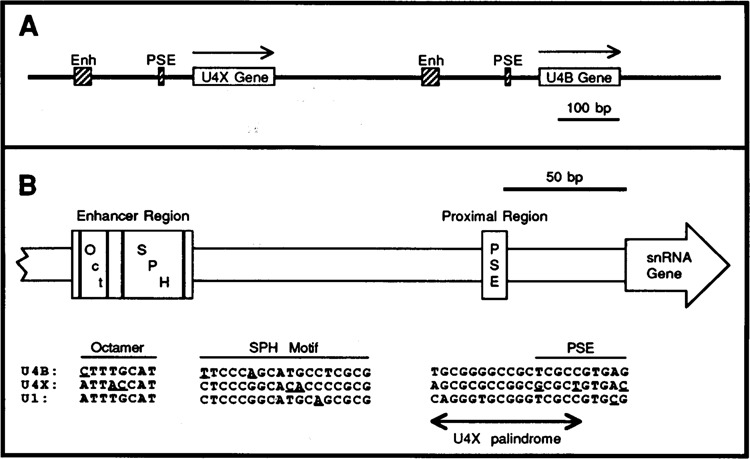
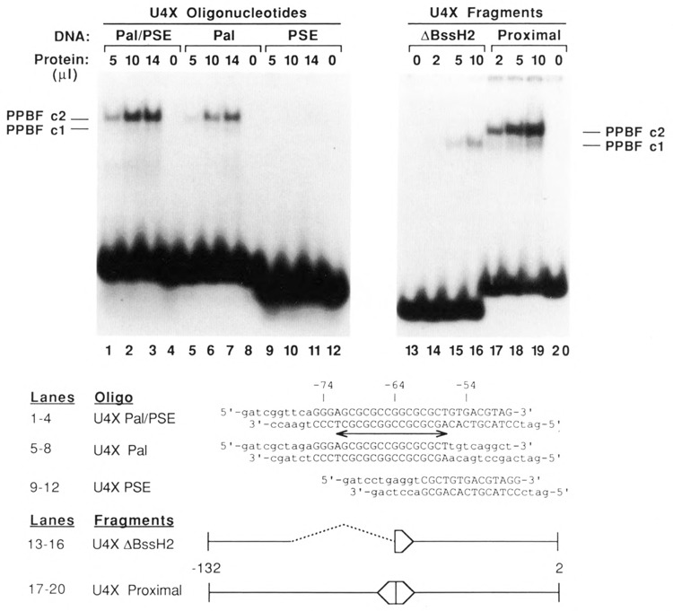
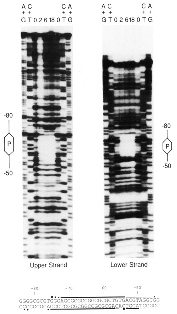
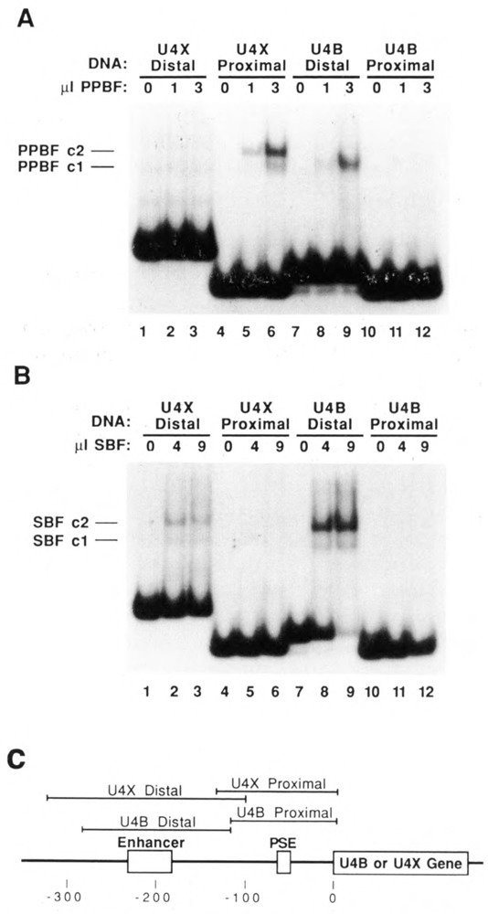
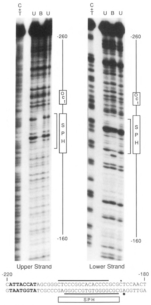
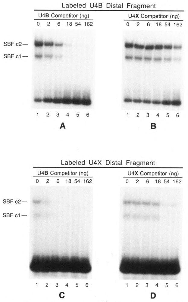
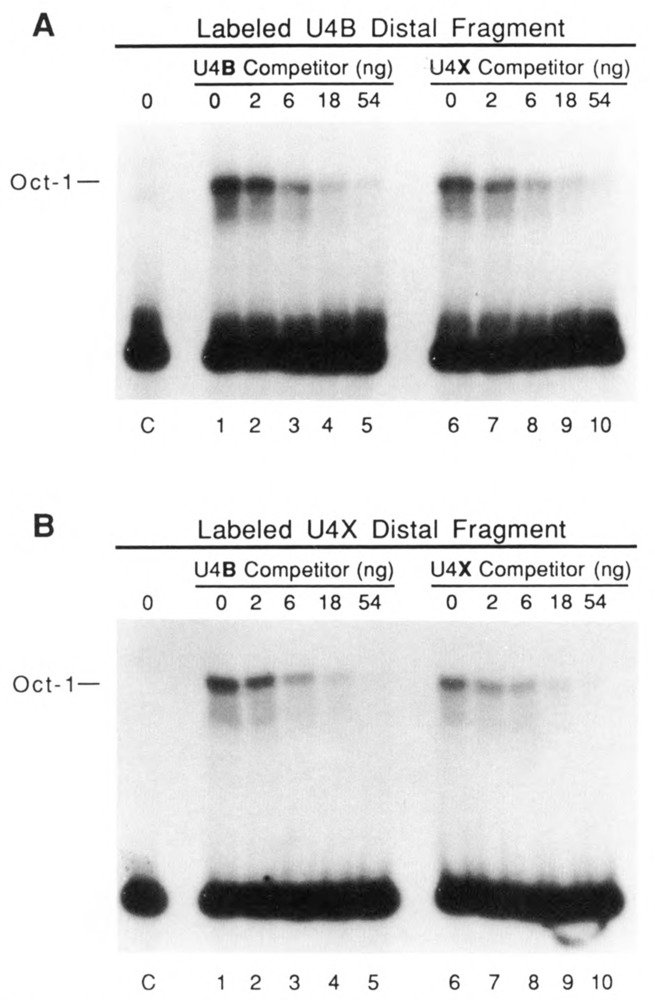
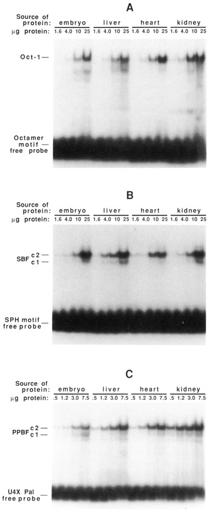
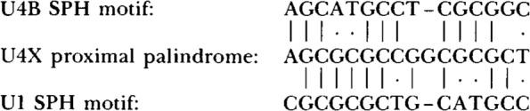
In the chicken genome there are two closely-linked genes, U4B and U4X, that code for different sequence variants of U4 small nuclear RNA (snRNA). Both genes are expressed with nearly equal efficiency in the early embryo, but U4X gene expression is specifically down-regulated relative to U4B as development proceeds. At the present time, little is known about the mechanisms that regulate differential expression of snRNA genes. We have now identified a novel chicken factor, PPBF, that binds sequence-specifically in vitro to the proximal regulatory region of the U4X gene, but not to the proximal region of the U4B gene. PPBF is itself regulated during development and may therefore be a key factor involved in differentially regulating U4X gene transcription relative to U4B. The U4X and U4B enhancers contain distinct sequence variants of two essential motifs (octamer and SPH). The Oct-1 transcription factor binds with similar affinities to both the U4X and U4B octamer motifs. However, a second essential snRNA enhancer-binding protein, SBF, has a 20- to 30-fold lower affinity for the SPH motif in the U4X enhancer than for the homologous SPH motif in the U4B enhancer. A potential role therefore exists for SBF, as well as PPBF, in the preferential down-regulation of the U4X RNA gene during chicken development.


Figures

Figure 1
Figure 1
A. Genomic organization of the chicken U4X and U4B snRNA genes. The genes are transcribed left-to-right, as indicated by the arrows. Cross-hatched boxes indicate the locations of the enhancer (Enh) and proximal sequence elements (PSE) conserved in the 5′ flanking DNA of vertebrate snRNA genes. B. Similarities and differences in the promoters of the chicken U4B, U4X, and U1 snRNA genes. The schematic diagram shows the locations of the PSE and of the octamer and SPH motifs that are conserved at nearly identical positions upstream of the U4B, U4X, and U1 genes. The sequence comparisons at the bottom of the figure show that among these three genes the regulatory motifs are quite similar but not identical in sequence. Underlines point out nucleotides in the octamer, SPH, and PSE motifs that are different in one sequence but conserved in the other two. The double-ended arrow indicates the location of a palindromic sequence unique to the U4X gene that partially overlaps the U4X PSE. The U1 sequences shown are from the Ul-52a gene (Earley et al., 1984). The U1 gene octamer and SPH motifs occur naturally in the opposite orientation compared to those in the U4 genes; therefore the U1 octamer and SPH motif sequences shown in the figure are from the template strand, whereas all other sequences are from the non-template strands.
Figure 2
Figure 2
Identification of PPBF, a factor that binds specifically to a palindromic sequence in the proximal regulatory region of the U4X gene. EMSAs were performed by incubating increasing amounts of affinity-purified factor, PPBF, with various synthetic oligonucleotides and restriction fragments that contained DNA sequences from the U4X gene proximal region. The sequences of the synthetic DNA oligonucleotides are shown below the autoradiograms. The U4X Pal/PSE oligonucleotide contained the complete PSE and overlapping palindromic sequence, whereas the U4X Pal and U4X PSE oligonucleotides contained complete sequences only for the palindrome or the PSE respectively. Wild-type nucleotides are shown in upper case, and the double-ended arrow indicates the position of the palindrome. The restriction fragment designated U4X proximal contained U4X sequences from position −132 in the 5′ flanking DNA to position 2 in the coding region of the gene. The U4X ΔBssH II fragment was similar, except that nucleotides from positions −105 to −64 had been deleted by BssH II digestion and re-ligation. Only DNA oligonucleotides or fragments that contained the intact palindromic sequence were able to form the upper complex, c2.
Figure 3
Figure 3
Footprint of PPBF on the U4X proximal region. DNA fragments end-labeled on either the upper or lower strand were incubated with 0, 2, 6, or 18 μl of affinity-purified PPBF (as indicated above the individual lanes), subjected to partial DNase I digestions, and analyzed in denaturing gels. A + G and C + T chemical sequencing reactions were run alongside as markers. The location of the palindromic sequence in each ladder is indicated by the hexagons labeled “P,” and the extent of DNA sequences from −50 to −80 (relative to the transcription initiation site) is delineated by the lines extending from the hexagons. (In the fourth lane of the left panel [2 μl PPBF], the bands are light due to over-digestion and partial loss of sample.) At the bottom of the figure, the nucleotide sequence of the U4X proximal regulatory region is shown, and the horizontal lines indicate the protected regions on the upper and lower strands. Thick lines denote strong protection, and thinner lines weaker protection. Dots indicate sites of enhanced DNase I cleavage induced by the binding of PPBF.
Figure 4
Figure 4
EMSAs demonstrating that PPBF and SBF have distinct DNA-binding specificities. A. Restriction fragments encompassing the U4X enhancer, U4X PSE, U4B enhancer, or U4B PSE were end-labeled and incubated with 0, 1, or 3 μl of affinity-purified PPBF (as indicated above each lane); the formation of protein-DNA complexes was analyzed by electrophoresis in a native polyacrylamide gel. B. The same four DNA fragments were incubated with 0, 4, or 9 μl of affinity-purified SBF, an snRNA gene enhancer-binding protein; complex formation was analyzed as in A. C. Diagram showing the regions encompassed by the U4X distal, U4X proximal, U4B distal, and U4B proximal restriction fragments used in the EMSA analyses presented in A and B.
Figure 5
Figure 5
Footprint of SBF on the U4X enhancer region. A restriction fragment encompassing the U4X enhancer region was labeled on either the upper or lower strand at approximately position −105, incubated with affinity-purified SBF, and partially digested with DNase I. The sample was loaded on a preparative EMSA gell and bands corresponding to bound (B) and unbound (U) DNA fragments were eluted and run in separate lanes of a sequencing gel. The lanes labeled C + T contained chemical sequencing reactions as markers. The region protected from DNase I digestion by bound SBF is indicated by brackets alongside the autoradiograms. The protected region is also denoted at the bottom of the figure by horizontal lines above and below the DNA sequence of the U4X enhancer region. Dots indicate sites of enhanced DNase I cleavage induced by the binding of SBF. The rectangular box labeled SPH points out the 18 bp of sequence similarity shared with the U4B and Ul SPH motifs. The nearby octamer sequence variant is shown in bold type.
Figure 6
Figure 6
EMSAs demonstrating the differential interaction of SBF with the U4B and U4X enhancers. Assays were performed by incubating 6 μl of affinity-purified SBF with a 32P-labeled DNA fragment encompassing either the U4B enhancer (A and B) or the U4X enhancer (C and D). Incubation mixtures also contained as specific competitor DNA increasing amounts of either the unlabeled U4B fragment (A and C) or the unlabeled U4X fragment (B and D). In each case, the U4B fragment was a 20- to 30-fold more effective competitor than the U4X fragment. The autoradiograms in C and D are 3-fold longer exposures than those shown in A and B. The U4B and U4X distal DNA fragments are diagrammed in Fig. 4C.
Figure 7
Figure 7
Oct1 factor has similar affinities for the U4B and U4X enhancers. EMSAs were performed similar to those in Figure 6, except that in vitro translated Oct1 factor was used in place of SBF. A and B show results obtained by using labeled DNA fragments from the U4B and U4X distal regions, respectively. Reactions loaded in lanes 1-5 (both panels) contained increasing amounts of the unlabeled U4B distal fragment as competitor, and reactions loaded in lanes 6–10 (both panels) contained increasing amounts of the U4X distal fragment as competitor. Lanes labeled C contained control reactions lacking competitor but incubated in translation extracts from which in vitro synthesized Oct1 mRNA had been omitted.
Figure 8
Figure 8
DNA-binding activities of Oct1, SBF, and PPBF as a function of development and tissue-type. Nuclear protein extracts were prepared from 8-day embryos and from liver, heart, and kidneys of chicks 3 weeks post-hatching. Increasing amounts of each unfractionated extract were incubated with 32P-labeled double-stranded oligonucleotides containing specific binding sites for Oct1 (A), SBF (B), or PPBF (C) and analyzed on EMSA gels. Oct1 and SBF DNA-binding activities appear relatively constant in the various tissues; the specific DNA-binding activity of PPBF is lowest in embryo, intermediate in liver and heart, and highest in kidney.
None

Similar articles

Cited by

References

    1. Dahlberg J. E. and Lund E. (1988), in Structure and Function of Major and Minor Small Nuclear Ribonucleoprotein Particles (Birnstiel M. L., ed.), Springer Verlag KG, Heidelberg, pp. 38–70.
    1. Earley J. M. III, Roebuck K. A., and Stumph W. E. (1984), Nucl Acids Res 12, 7411–7421. - PMC - PubMed
    1. Forbes D. J., Kirschner M. W., Caput D., Dahlberg J. E., and Lund E. (1984), Cell 38, 681–689. - PubMed
    1. Gunderson S. I., Knuth M. W., and Burgess R. R. (1990), Genes Dev 4, 2048–2060. - PubMed
    1. Hernandez N. and Lucito R. (1988), EMBO J 7, 3125–3134. - PMC - PubMed

Publication types

MeSH terms

LinkOut - more resources